Jennifer Lake's Blog

May 14, 2023

Corona the Crow, Part Four

*

Corona the Crow, Part One https://jenniferlake.wordpress.com/2022/06/10/corona-the-crow/

Part Two https://jenniferlake.wordpress.com/2022/12/06/corona-the-crow-part-two/

Part Three https://jenniferlake.wordpress.com/2023/01/02/corona-the-crow-part-three/

PART FOUR of the Crow is going to focus on corona as empirical technology, particularly as a medical paradigm of being taken by the head — physically, psychologically and socially. Hopefully, as a reader, the preceding parts of this series have adequately demonstrated that Corona the Crow stems from ancient ideas about the human mind. Brain anatomy is loaded with crow words; cortex, cerebrum, and claustrum, for example, nested under your cranium. “Psi,” the crow-foot (or fish spear) letter from the Greek alphabet, is a symbol for the mind, adopted by societies of medicalized psychology.  The word ‘medical’ is itself an English latecomer, elaborated from the Latin word for ‘mediate’—medlare, to meddle– and the older Middle English word ‘medic’ which defined leguminous food plants like beans, subject to plantation methods in the birth of civilizations. A plant medic, according to my dictionary, has a distinctive structure of trifoliate leaves symbolized by example as the stylized Fleur-de-Lis: “fleur-de-lis, (French: “lily flower”) , also spelled fleur-de-lys, also called flower-de-luce…much used in ornamentation and, particularly, in heraldry, long associated with the French crown…” as a gift from the Virgin Mary, no less, who maintains her original status with me as a transplanted sea-bird. https://www.britannica.com/topic/fleur-de-lis .

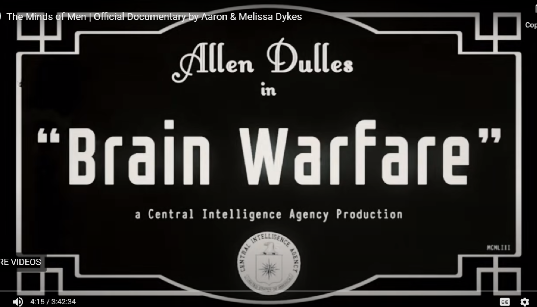
Luce, of course, is the seemingly familiar root word meaning ‘light,’ but its actual Middle French/English origins identify it with a fish (the pike) as it remains, representing a large family of freshwater specimens. https://www.dictionary.com/browse/luce ; https://www.britannica.com/animal/pike-fish. One can digress to Fisher Kings, Merovingian ‘mermaids’, holey (‘cro’) blood and the divine Right to Rule within our Clockwork of Time and Water, but I would sooner get on with Mind Control in its variable and loathsome permutations. That is what Corona is about.  Note in the 2018 documentary, The Minds of Men, this screen capture (minute 4) of the 1953 CIA-made film “Brain Warfare” starring Allen Dulles.

*

As Part Four progresses, I’ll be referencing The Minds of Men ( by Aaron and Melissa Dykes, 3hrs 43min, https://www.youtube.com/watch?v=LQucESRF3Sg )–noting the filmmakers own emphasis on MK-Ultra Subproject 119. The gematria (numerical letter) value of 119 is also the sum of the words Crow (=59) plus Raven (=60), mentioned in Parts Two and Three along with a lot of other ‘1’s and ‘9’s.

If you wanted to be a fisher of luce men, so to speak, it would help to have a proper boat and a strong net …so, let’s recap the definition of corona from Part I:

“Derived from Greek κορώνη (korone) meaning ‘crow’ … In Greek mythology, Coronis (/kɒˈrəʊnɪs/; Greek: Κορωνίς, translit. Korōnís) is a Thessalian princess and a lover of Apollo… By Apollo she became the mother of Asclepius,[2][3][4][5] the Greek god of medicine…”

Aesculapius and others like him were … “sprung from an egg”… “where a serpent in an egg is introduced as a true emblem of this deity… the emblem Coronis must sometimes have had a reference to the same…yet…Coronis was certainly a name given by the poets and mythologists to a particular machine: the same in which the gods are supposed to have been enclosed and to have suffered a temporary death. Coronis is accordingly said to have been a vessel or float… By Coronis is signified a ship…”

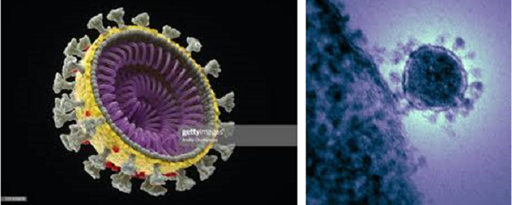
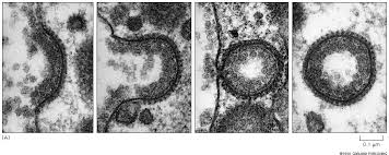
…………… here’s a ‘serpent in an egg’………shipping………FYI, these are official images of coronavirus presented by the mass media.

*

In this recent April 30, 2023 interview with Maria Zeee, Dr. Stephanie Seneff proposes a mechanism for shipping ‘virus’ particles from the Covid-19 injection site in the arm along nerve fibers to the brain, proofing on the symptoms of fatal neurological diseases like Mad Cow, or Creutzfeldt-Jakob Disease (CJD) in humans, and ALS (Amyotrophic Lateral Sclerosis, or ‘Lou Gehrig’s’) seen in post-‘vax’ injuries during the early months of 2021.  The publishing source of these fatal effects came from an April 2021 paper co-authored by Luc Montagnier, mentioned at minute 36 by Dr. Seneff: “They know that it can cause a severe [fast-acting] form of Creutzfeldt-Jakob Disease, which is exactly what we’re seeing in those 26 patients that Luc Montagnier wrote about in the paper that got published after he died.”

Here’s the shipping statement in the transcript at minute 24–[Seneff:] “…That spike protein—it’s amazing because it looks like it’s been designed to be a bioweapon and it has so many toxic aspects to it that it’s disturbing to think [it can cause] your muscles to keep spewing out spike proteins and throwing it into exosomes and distributing it throughout your body… I want to get into this issue of distributing it along the nerve pathways, what I’ve come to believe, as a whole other topic [in addition to vaccine-carrying food plants] that I think I’ve finally gotten right…  [minute 27–] So, here’s the story: The muscle cells take up nanoparticles, which look like LDL particles and the muscles have LDL receptors so they take up the particles, open [them] up and start making spike protein…, spitting it out as exosomes and releasing them into the synapse. So now, the muscles which are enervated by motor neurons…which have super long thread[s] from the spinal cord –axons, well, those axons are at the synapse to cause muscles to contract…[and] in that synapse you have all those [muscle cell-made] exosomes…releasing spike protein…and mRNA…and the axon will take up those exosomes [even though] it won’t take up the nanoparticles [because axons] don’t have LDL receptors…but they will take up exosomes. So now, [axons are] taking up exosomes and there are active transport proteins –there’s a molecular motor called dynein which is in the axon—that specializes in shipping stuff up the axon through the soma of the cell which is in the spinal cord… [and the exosomes get packaged into what are called endosomes] ..and the exosomes survive inside the endosome —the whole package gets shipped up to the core of the nerve cell that’s controlling muscle movement…and up the spinal cord to the brainstem.” https://zeeemedia.com/interview/uncensored-dr-stephanie-seneff-can-mrna-bypass-our-stomach-acid-if-they-put-it-in-our-food/

For the moment, Dr. Seneff’s description of nerve cells shipping loaded particle packages “up your tree” sounds reminiscent of the Queen’s Platinum Jubilee—yes! the T-Mobile Crow Show of last June with the overt lighting display of traveling DNA (linked in Part One). But there are many more correspondences related to this interview besides infection mechanics.  In 1953 when the CIA issued the Brain Warfare film, the poliovaccine was initialized at small-scale human testing– testing whether subjects could survive the shots. The next year in ’54, the test expanded to 1.8 million schoolchildren followed in ’55 by nationwide mass vaccination. In some of the coverage areas polio stats skyrocketed from 400% to 700% over pre-vaccine case numbers.

Here’s a brief excerpt posted at the Polio Forever blog from the book The Polio Paradox on the subject of latent, later-in-life post-polio, by Richard L. Bruno:

Chapter Three “The Guided Missile”…[p20] quoting researcher David Bodian: “The average number of spinal cord motor neurons that are destroyed by the poliovirus is almost 50%. However, nonparalytic polio may be associated with severe neuron damage in the spinal cord. What is more, some with nonparalytic poliomyelitis do not have any damage in the spinal cord but have characteristic damage in the brain, which is more extensive than in some who did have paralysis. All available evidence shows conclusively that every case of polio exhibits damage in the brain. The poliovirus is capable of producing an encephalitis, with or without symptoms, in the absence of any damage to the spinal cord. As far as the pathologist is concerned all cases of polio are encephalitic”. [p21] It’s amazing that something so small can do so much damage…But the poliovirus doesn’t attach to and damage just any cell. It is a ‘guided missile’ that does one thing: seek out, damage, and destroy the neurons that “activate” you –the ones that activate your brain and muscles. The poliovirus is the perfect human “Off switch”… https://polioforever.wordpress.com/the-polio-paradox/

*

Polio is fundamental to the fields of Psychiatry, Psychology and Neurology:

On the record, the disease we came to know as ‘polio’ in mid-century America arose with industrialization and occupational exposure to toxic metals and chemicals –prior to the 1850s, the ‘flaccid paralysis’ of acute illness was rare enough to be medically unknown. Small epidemics (10-20 cases) were first observed and recorded among iron-and-steel mill workers in Norway and Sweden. In the following decades, the spotty appearance of polio-like disability began to affect numerous industrial workers across the range of chemical users. One famous polio case in particular, “the French patient” youth who worked in the leather dye-solution vats at a tannery, virtually assured the ascending reputation of neurologist Jean-Martin Charcot (1825-1893) who classified several of the neurodegenerative diseases of the 20th century, among them ALS (Lou Gehrig’s) and Multiple Sclerosis. Polio, by historical measure, gave rise to the academic fields of Neurology and Psychology, a two-way schism that developed around the 1870s and onwards from the study of ‘neurasthenics’. In our corona Mind Control context, the 1901 founding of the Rockefeller Institute for Medical Research (RIMR, renamed Rockefeller University) stands as the first milestone marker. RIMR’s establishment mission statement was to “pasteurize milk and study polio.” Almost no doctors in the U.S. had ever heard of poliomyelitis. In 1953, again, with the Salk vaccine in human testing and on the near horizon, the medical establishment altered polio’s diagnostic criteria. Even Franklin Delano Roosevelt posthumously had his polio changed to Guillain-Barre Syndrome, a top ranking ‘side effect’ of Covid-19 injections. As of today, I’ve learned that the most salient facts about polio are (1) that radiation exposure is a stand-alone cause, most likely by x-rays from the 1890s forward, and (2) the ‘germ’ was unknown until the mid-1950s following the vaccine and is most likely sourced from plants.

 Read more about the far-reaching Rockefeller Institute https://polioforever.wordpress.com/the-rockefeller-institute/

An update due to the polio pages which were posted back in 2011 requires another look to our crow friends who were felled in 1999 from a mosquito-borne contagion said to be West Nile Virus:  “West Nile virus hit American Crows particularly hard. When the disease first appeared in New York City, in summer 1999, nearly 5,500 crows died in 4 months. Tests suggested the disease was 100-percent fatal to crows. Many other species, from jays and magpies to gulls and chickadees, also proved susceptible. Millions of birds died as West Nile swept across the continent in just five years…” [2010 issue of BirdScope]” https://www.allaboutbirds.org/news/counting-crows-the-impact-of-the-west-nile-virus/#  

More recently than 2010, the WHO has added this highlight (in bold): “West Nile Virus (WNV) was first isolated in a woman in the West Nile district of Uganda in 1937. It was identified in birds (crows and columbiformes) in Nile delta region in 1953. Before 1997 WNV was not considered pathogenic for birds, but at that time in Israel a more virulent strain caused the death of different bird species presenting signs of encephalitis and paralysis… [In humans, the] symptoms of severe disease (called neuroinvasive disease, such as West Nile encephalitis or meningitis, or West Nile poliomyelitis) include headache, high fever, neck stiffness, stupor, disorientation, coma, tremors, convulsions, muscle weakness and paralysis… Birds are the reservoir hosts…” https://www.who.int/news-room/fact-sheets/detail/west-nile-virus ; the 1953 intel appears to be new.

*

Author of the 2017 book The Invisible Rainbow, Arthur Firstenberg, wrote, “In 1982 I was in my final year of medical school… One day I collapsed and was unable to get up… It wasn’t a heart attack, but it was six months before I could walk up a flight of stairs without becoming short of breath… I gradually found that I was being effectively disabled by my society. Having stumbled upon an obviously well-kept secret, I researched the world literature on bioelectromagnetics… together with other members of a support group I had found how best to live with my disability… But in July 1996, to my dismay, I learned that an innovation was coming to my city which threatened to make it impossible to avoid exposure any more… [On] November 14, 1996, Omnipoint, New York City’s first digital provider..open[ed] for business… According to health authorities, an early flu hit New York City –but not Boston and not Philadelphia– on about 15 November… I learned that in February 1996, Congress had passed a law prohibiting local governments from denying permits for cell phone antennae because of environmental concerns… I also learned that the FCC had just issued regulations setting public exposure limits for microwave radiation at levels..ten thousand times higher than levels..causing reports of illness from all over the world.” http://www.whale.to/b/elfb.html

Omnipoint, est.1987; “before Omnipoint, the use of encryption technology was mostly limited to  military communications. Omnipoint is the first and only company in the New York metropolitan area to use GSM – Global System for Mobile communications.” http://www.schneier.com/cmea-omnipoint.html

There is enough circumstantial evidence to suggest that Omnipoint’s Global System for Mobile (GSM) signal operations were among the mechanisms spreading West Nile and much of it is in the polio literature.

 “The GSM standard was accepted in the United States in 1995. GSM-1900 cellular systems have been operating in the US since 1996.” http://inventors.about.com/library/weekly/aa072199.htm

   Omnipoint’s “Personal Communications Systems, PCS..[was] a newer class of wireless..services..authorized by the FCC [which] use a different radio frequency, the 1.9 GHz band, than cellular phones and generally use all-digital technology for transmission and reception… In 1994, the FCC announced it was allocating spectrum specifically for PCS..” http://inventors.about.com/library/weekly/aa072199.htm

“PCS differs from existing cellular and SMR in three basic ways: frequency, spectrum and geographic division. PCS networks operates in a higher-frequency bank (1850-1990 MHz) compared to the cellular and SMR frequency (800-900 MHz). The higher frequency band for PCS networks requires a greater number of total cell sites to cover the same geographic area as existing cellular networks… http://www.hotstocked.com/companies/n/northeast-digital-networks-inc-GSMI-description-68560.html

Dec.1997–“In wireless, it is often said, coverage is king. Omnipoint has had its fair share of cell siting hassles. To get over objections, the carrier has placed its transceivers inside gargoyles on the facades of landmark buildings in Manhattan..http://www.rcrwireless.com/article/19971229/sub/rcrs-1997-person-of-the-year-george-schmitt-omnipoints-george-schmitt-burns-the-midnight-oil-to-build-pcs-firm/

New York’s Office of Emergency Management (OEM) system could also be to blame: NYC “OEM frequencies transmit in a narrow range at 147 MHz.” http://www.dhses.ny.gov/oem/radiological/

…human neuroblastoma cells were exposed to electromagnetic radiation at 147 MHz, amplitude-modulated (AM) at 16 Hz… Enhanced efflux at 0.05 W/kg peaked at the 13-16 Hz and at the 57.5-60 Hz modulation ranges…These results confirm that…radiofrequency radiation can induce responses in cells of nervous tissue origin from widely different animal species, including humans…” http://www.scribd.com/doc/34980786/RF-Induced-Calcium-Ion-Efflux-Enhancement-From-Neuroblastoma

Dr [Ross] Adey demonstrated how a 147-megahertz (MHz) field, which at tissue level had  an intensity of 0.8 milliwatts per square centimetre, caused an efflux or  release of calcium ions from the irradiated brain tissue…” http://www.whale.to/b/adey.html

In 2002 Omnipoint became T-Mobile https://www.reference.com/business-finance/omnipoint-communications-new-york-9e34507adf4eba25 ; Dr. Adey is one of several researchers in the 50s and 60s known to have participated in MK-Ultra Subproject 119.

*

MKULTRA Subproject 119

 “Activation of human behavior by remote means.”

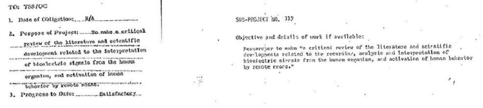
” 2. Purpose of Project: To make a critical review of the literature and scientific development related to the interpretation of bioelectric signals from the human organism, and activation of human behavior by remote means. 3. Progress to Date: Satisfactory.”

sources:

https://archive.org/details/DOC_0000017376/page/n1/mode/2up

https://www.illuminatirex.com/list-of-mkultra-subprojects/

_______________________________

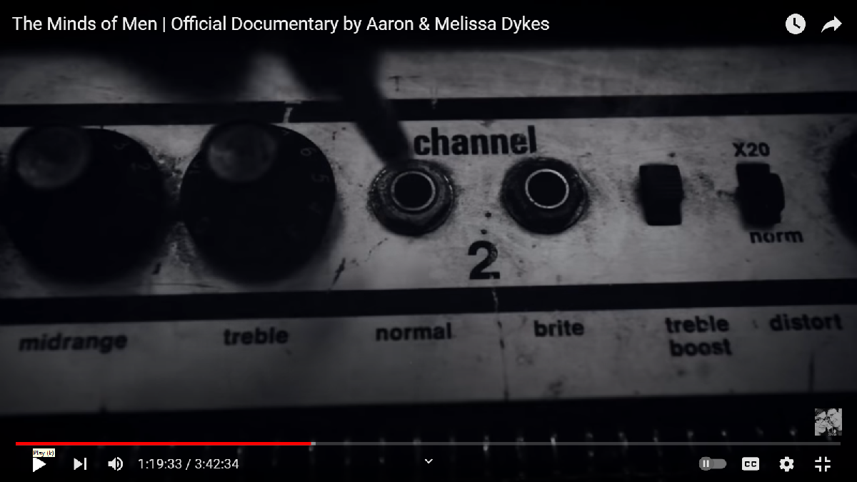
Among the technical successors to MKULTRA’s Subproject 119 is optogenetics which in recent years has gone wireless thanks to a light-sensitive protein from algae called ChRmine (pronounced KAR-meen) described here by Karl Deisseroth, a leading researcher at Stanford. Listen through minute 25 https://www.youtube.com/watch?v=Ls07NZRJp6E ; It’s no longer necessary to cable a light source into the brain.  Deisseroth says of ChRmine: “you don’t have to bring in light powerbut you still have to bring in the gene.

min24: “…[Behaviors are] assembled from parts, just like anxiety, and are tunable and can be brought down to the physical elemental components using optogenetics. So that’s pretty interesting, but then ChRmine changes the [?’sass’?]. We do like the fiber optic method—we can bring in light to a particular region but the extreme light sensitivity of this new channelrhodopsin means we can even do noninvasive light delivery. Here’s an example of that: We can have the fiber optic completely outside the brain of the animal and we can target deep structures like the ventral-tegmental-area dopamine neurons. Using an electrode we can track how well we’re doing and we can actually drive these neurons, drive the action potentials of these neurons, without brain-penetrating hardware at all. In fact, you can use non-injection-based tricks to get the gene in too—we [already] have ways of getting [the genes] into the brain with an intravenous injection, and this was developed by a friend, post-doc grad student of mine named Vivianna Gradinaru…now at Caltech. So, you can do completely noninvasive deep fast control of neurons now. And that raises all kinds of questions…”

*

…and my question is: Were Covid vaccines shipping mRNA-making optogenetic ‘light’ proteins, and will there be more of it in future mRNA vaccines? Some potential visual clues about future mind control technology in the Dykes documentary seem very suggestive despite appearances of past history. The opsin proteins at issue are called channelrhodopsin 2, or ‘channel 2’ images, shown several times in the video but shown first at 1:19 (1hr 19 minutes plus 33 seconds precisely). A clue? Do they teach that in film school? The significance of movie timecodes and numbers is covered in Part Two where I had my initial Back to the Future moment with the Crow.

Bringing in the genes to optogentically modulate humans, they tell us, requires transfection:

“So, the technology is relatively simple. You take DNA from a microorganism, Chlamydomonas for example, and connect it with a promoter region from the cell of interest, pack it into a virus, inject the virus into the brain, and then you wait… and study the behavior…” —minute 29, from speaker Peter Hegemann, “Optogenetics: Illuminating the Path toward Causal Neuroscience”, a Harvard prize-giving presentation recorded in 2019 https://www.youtube.com/watch?v=hOKLMjuwlLo

Now that we have a working model of ‘muscle-to-axon’ infection mechanics on how to “inject the virus into the brain” let’s check back in with our White Raven researchers:

[Sep15, 2021]  “Biochemist Katalin Karikó and her colleague Drew Weissman were recently awarded a $3-million Breakthrough Prize for their work… Karikó, who is now senior vice president and head of RNA protein replacement therapies at BioNTech (the company that co-developed a COVID vaccine with Pfizer), and Weissman, a professor of vaccine research at the University of Pennsylvania’s Perelman School of Medicine, have just been awarded…for their work on modifying the genetic molecule RNA…  Karikó spent years on this research despite skepticism and a lack of funding. Ultimately, however, her efforts paid off…  Weissman and his colleagues had been working on an mRNA vaccine for influenza when word spread of a mysterious pathogen causing pneumonia in people in Wuhan, China, in late 2019. Weissman quickly realized this virus was a perfect candidate for an mRNA vaccine, and Pfizer-BioNTech and Moderna soon pivoted to work on one. The rest is history… [said] KARIKÓ: ‘From 2018 we had worked with Pfizer to develop a vaccine for influenza. And we were already ready to start a clinical trial for that. But switching over to COVID, it was just a technical thing. And so it was already ready. If the pandemic had happened 20 years ago, you would need to have, physically, in your hands, a piece of the virus. So that would be a big delay. But commercial gene synthesis started about 20 years ago. Now you can just order a gene. You order DNA, and then you insert it into a [typically circular molecule of DNA called a] plasmid, and then you make RNA. But making the nanoparticle to deliver the mRNA is kind of challenging.’ [question:]The lipid nanoparticles were a key part of the technology to make it useful for vaccines, right? KARIKÓ: ‘In my view, yes. The lipid nanoparticle protects the mRNA outside the cell because, in the blood and everywhere, there are a lot of human enzymes that can degrade the RNA. Second, it helps it to enter because the [muscle] cell will pick up the particle. And then it is in the endosome [a membrane-bound compartment] in the immune cells, and then this lipid nanoparticle helps escape from the endosome to the cytoplasm [the solution inside cells] so the protein can be made. It is a very smart particle.’ ” https://www.scientificamerican.com/article/an-mrna-pioneer-discusses-how-her-work-led-to-the-covid-vaccines/

*

Influenza mRNA injections “already ready”:

From Redacted News, May 17, 2023: “It’s STARTING—the NEXT pandemic is here & children are the target says Bill Gates” [17 minutes]: “…”it’s coming and right on cue the UK government today, this morning, announced it’s testing for a new avian flu outbreak… watch [this UK news clip announcer]…”it regards bird flu…and a real concern about it being passed from wild birds to housed poultry…and now…in two people…” https://www.youtube.com/watch?v=8PqlzOVfciQ

      Bird Flu, H1N1 –or “Have 1, Need One World or None” and counting:

H1N1 (2009)– a visual match for coronavirus

..or…coming to a theater near you… H5N1 Bird Flu [posted May 21, 2023] “…scientists have long considered H5N1 to have pandemic potential. The U.S. has a stockpile of H5N1 flu shots in case such a crisis arises, but…[t]he shots have only been administered in trials and were derived from strains that circulated in 2004 and 2005… [It’s] suggested that a bird flu pandemic would most likely start as a small outbreak among poultry workers or swine workers, since pigs can pass the virus from birds to humans…  Moderna said that later this year, it expects to begin clinical testing of an mRNA vaccine specific to the strain now circulating in birds, called 2.3.4.4b… a universal flu vaccine, which would target a wide variety of flu strains, could also provide cross-protection against whatever version of bird flu one day finds its way into humans. Several such vaccines are being developed… The National Institutes of Health announced this month that it has started testing a universal mRNA flu vaccine among 50 volunteers.” https://www.nbcnews.com/health/health-news/bird-flu-spreads-people-existing-vaccines-may-inadequate-rcna72973

H5N1

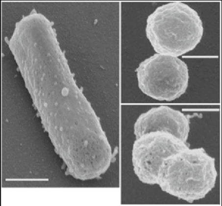
H5N1 labeled here, showing both ‘rod and sphere’ forms apparently, is looking very much like Wikipedia’s pictures of Halobacterium, the light-activated ‘opsin’  protein carrier discovered in the 1960s as the first optogenetic tool in the toolkit. https://en.wikipedia.org/wiki/Halobacterium_salinarum ; https://microbewiki.kenyon.edu/index.php?title=Halophiles&redirect=no

                        _______________________________________

“The greatest weapon of the oppressor is the mind of the oppressed” –Steven Biko

                        _______________________________________

The Disease Continuum is the title of a post I put up in 2011, having the negative distinction of being among the most ignored articles on the blog. Was it timing? The title?—maybe I was preparing for yet unnamed Covid. (#98 in BLOG INDEX)  I promised a serial, but never brought it back under the ‘continuum’ heading:

“In general terms, the mater materia of the Disease Continuum compares to the elements of the ancients [air, water, earth, fire]; four fundamental essences from which it is composed. By disease names they are influenza, polio, cancer and AIDS andtogether they forge a Ring of Power in the kingdom of Public Health. Common to the four elements of the Continuum is the eugenical substrate on which they emerge in history; they are timely, political, and as inevitable as the science and industry that sustains them. It should interest us that they are characterized as viral and not the foreign biological intruders we suppose. ‘We have travelled a long way from the mysterious filtrable infective particle of…years ago… [W]e have even the evidence that…portions of certain…viruses can be dissociated and later recombined to form a reconstituted infective particle… Clearly discoveries of this sort are providing the basis for an understanding of the host-virus relationship… For virus multiplication is after all a special case of protein biosynthesis… We seem thus to have reached a point at which biochemical and biophysical studies of viruses have really come into their own and offer the greatest prospects of advance.’ –Sir Charles Harington, March 1956, Ciba Foundation Symposium at the National Institute for Medical Research (NIMR), Mill Hill London [ref. The Nature of Viruses, 1956, Little Brown & Co.]…

… Work on polioviruses helped to prove that intestinal “Enteroviruses can infect all tissues of the human body. The tropism of each virus for certain tissues is not well understood…”. Reconstituting pathogens in the form of gut bacteria and viruses was learned early. Simon Flexner designed experiments in 1897 to alter the properties of harvested human colon bacilli and turn them virulent several years before he became the director of the Rockefeller Institute for Medical Research. Flexner’s cadavers in 1890s Baltimore, taken to the labs of the newly medicalized Johns Hopkins University, were mostly victims of pneumonia and influenza, a ready surfeit of bodies that littered the northern port cities of industrial America.  https://jenniferlake.wordpress.com/2009/07/30/enter-o-viruses/

[and]… we should remember the words of H.R. Shepherd, 1993 founding chairman of the Sabin Vaccine Institute:  ‘Vaccines are the most powerful tool available to equalize the health of human beings in every corner of the world. Enlightened leaders understand the power of vaccines to help bring peace and opportunity to the most troubled places…’ http://polioforever.wordpress.com/sabin-vaccine-institute/

   No story of great or worldly achievement in the 20th century seems complete or soluble without a reconciliation to public medicine. It electrifies the most compelling events of our time like the JFK assassination. https://jenniferlake.wordpress.com/2011/11/06/the-jfk-conspiracy-con/

   Edward Mandell House, the intimate alter-ego and adviser to Woodrow Wilson, was reputed to have said (prior to WWI), ‘Very soon, every American will be required to register their biological property in a national system designed to keep track of the people… They will be our chattel… stripped of their rights and given a commercial value…‘ “

[read all] https://jenniferlake.wordpress.com/2011/12/07/the-disease-continuum/

*

[minute 1:21:56, The Minds of Men:] “When you run circles around the military’s technologically driven future, the nerves of the Army and Navy and Air Force, the communication networks of signals and intercepts…[and] when you run circles around the whole of human society and its various cycles of influence and modification…[and] the problem of the machine and man himself—once you go around and around the problem of feedback and cycles of behavior,…perception and reality, in the end the whole of Man can be controlled through his dependence on his environment.”

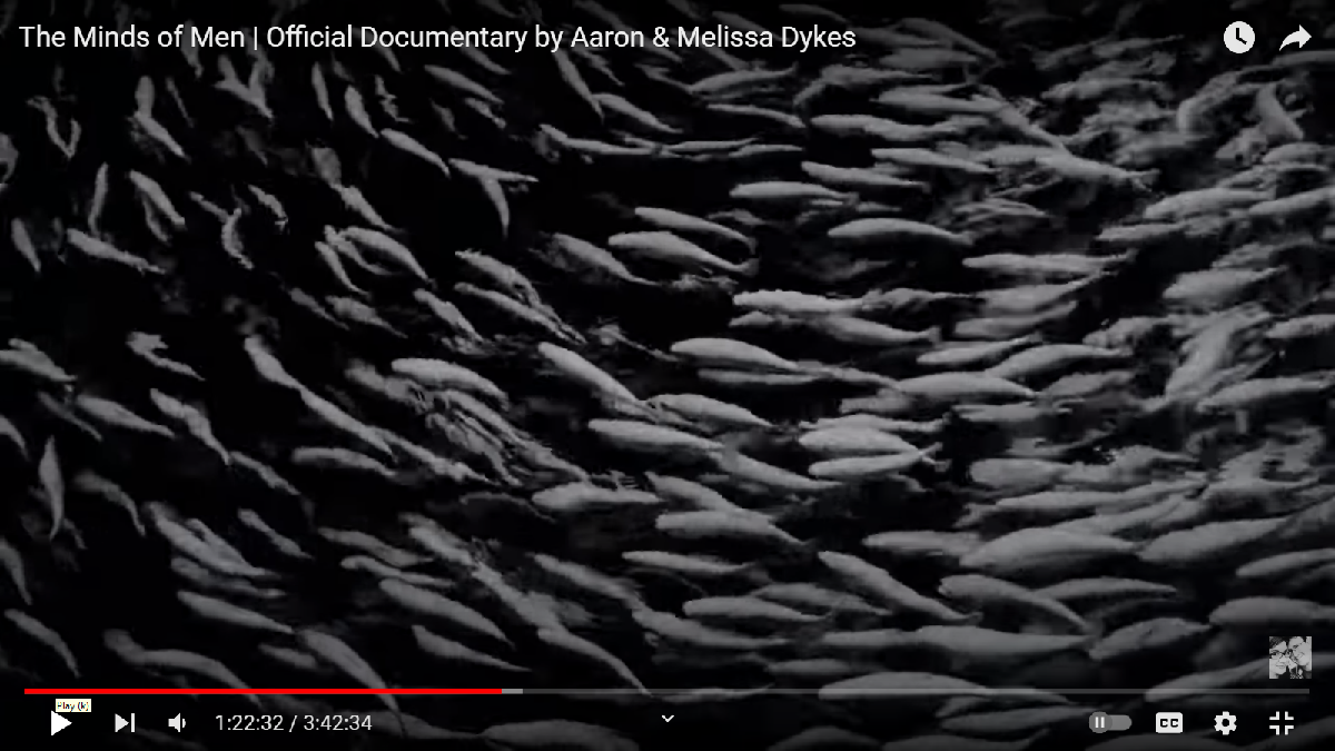
Schooling fish, waves on water…  appropos to the NET: (min 1:24 narration:) “Many of the cybernetics members [of the Macy Conferences] were now talking literally about constructing an artificial matrix… As Alex[ander] Bavelas of MIT noted in his group dynamics study, ‘We are attempting now to describe the operation of a net…as a matrix…’  (min1:09 narration:) Macy was meshing the power of language communication and the symbolic exchange of information with the biological modes of control…[over] the organization of the mind which might one day be conquered one neuron at a time.”(from The Minds of Men, part 2 Cybernetics minute 40 to 1hr38)

Indeed. Optogenetics today. Quoting Ed Boyden: “I also want to talk about how different technologies might fit together in this grand quest to understand the brain… I think we need to think about an integrated toolset…that lets us control the brain…[and] watch the brain in action and interestingly, tools in that nature are emerging from the optogenetic toolset itself… And as much as people have brought electrodes into the brain for over a century, you can bring in optical fibers or other kinds of optical devices. The next question is do you need to make a molecule that converts light to electricity or do you find it?…[We] found…a member of this [proton-pump] family [of organisms] from the archaerhodopsin class [that] if you genetically express it in neurons and then shine green or yellow light, will powerfully pump protons out of neurons and silence their activity [called hyperpolarization]… and you can shine blue light on neurons and let positive charge in and you can activate them, letting you investigate what those neurons are sufficient to trigger.” [called depolarization, or activation]–Edward Boyden, minutes 1:08 to 1:14, “Optogenetics Illuminating the Path…” https://www.youtube.com/watch?v=hOKLMjuwlLo/ ; Archaerhodopsin-3 came from a species of fungus called Leptosphaeria maculans that had a light-driven proton pump, “and we got the gene that we nicknamed ‘Mac’ for short and we found that it was also able to silence neural activity” –like an ON/OFF switch—minute 1:16 ibid.

“Mutants of Archaerhodopsin-3 (AR3) are widely used as tools in optogenetics for neuroscience research…” https://en.wikipedia.org/wiki/Archaerhodopsin

*

“Leptosphaeria maculans is the most damaging pathogen of Brassica napus [canola],which is used as a feed source for livestock and for its rapeseed oil… As a bioengineering innovation, in 2010 it was shown that a light-driven protein from L. maculans could be used to mediate, alongside earlier reagents, multi-color silencing of neurons in the mammalian nervous system.” https://en.wikipedia.org/wiki/Leptosphaeria_maculans

*

” ‘Optogenetics borrows these light-sensitive proteins from plants and uses them in animal cells,’ said UI professor Kai Zhang, who developed the optogenetic tools for controlling mitochondria and lysosomes with blue light. ‘By attaching such proteins to organelles, one can use light to control the interaction between them, such as mitochondria and lysosomes shown in this work.’  … Further research from Zhang’s lab will include developing new optogenetic systems that work with different colors of light, including green, red, and infrared, since a longer wavelength will be needed to penetrate human tissue.
‘We would like to further expand the toolbox by introducing multicolor optogenetic systems to give us multiple ways to control how organelles behave and interact,’ Zhang said. ‘For instance, one color makes organelles come together, while the other color forces them apart. This way, we can precisely control their interactions.’ The team hopes to progress from using human stem cells for its research to testing the efficacy of its technique in animal models, as a step toward eventually testing the technique in humans through clinical trials. Diao said that other research groups are studying the use of magnetic fields and acoustic vibrations to accomplish results similar to the team’s light-based technique.https://www.photonics.com/Articles/Optogenetic_Tools_Restore_Cell_Function_with_Blue/a68295

And, of course, it’s not just food plant fungi yielding up more and more ‘light’ proteins as the haloarchaea prove:   “Traditionally, Archaea were isolated from extreme environments where they are dominant, but recently they have been detected in nearly all environments examined using culture-independent techniques. Methanogenic Archaea are present in the mammalian gut and oral cavities… halophilic Archaea, such as Halobacterium, were isolated from salted protein sources such as fish sauces and animal hides.” https://www.sciencedirect.com/science/article/abs/pii/B9780123739445001085

“Haloarchaea is …mostly predominant in the gut of people who live in Korea. Haloarchaea are most abundant in Koreans guts rather than methanogens due to their saltier diets. This also shows that the archaeome in the human gut can vary drastically depending on region and what is eaten…” https://en.wikipedia.org/wiki/Haloarchaea ; Confirming the dominant presence of haloarchaea in Koreans is this :[August 04, 2020]… “We surveyed the archaeome of faecal samples collected from 897 East Asian subjects living in South Korea. In total, 42.47% faecal samples were positive for archaeal colonization… We observed extensive colonization of haloarchaea (95.54%) in the archaea-positive faecal samples… Haloarchaea were relatively more abundant than methanogens in some samples…” https://microbiomejournal.biomedcentral.com/articles/10.1186/s40168-020-00894-x/

Strange it seems, but information like this adds to my speculation that the Halloween “Seoul Crush” reported on 10-29-22 was a Li-Fi event (see the number play at the end of Crow Part Two—“examples of 119s”)

[same 2020 paper on Koreans archaeome:]“Information regarding the community-based genetic and functional traits of archaea in animal habitats is scarce. We have previously reported occurrences of diverse members of extremely halophilic archaea (haloarchaea) in avian plumage** [13], as well as in food samples such as salt-fermented seafood…” https://www.ncbi.nlm.nih.gov/pmc/articles/PMC7409454/ ; **“Flamingoes (Phoenicopterus spp.) whose plumage displays elegant colors, inhabit warm regions close to the ocean throughout the world… In this study, viable red-colored archaeal strains classified as extremely halophilic archaea (i.e., haloarchaea) and belonging to the genera Halococcus and Halogeometricum were isolated from the plumage of flamingoes in captivity… To the best of our knowledge, this is the first report of viable extremophilic archaea in avian plumage…”   https://pubmed.ncbi.nlm.nih.gov/26553382/

(The flamingo Fire Bird, below, on display in the last scene of the London 2012 Olympic closing ceremony)

In you, on you, and around you, Halophiles: Salt of the Earth…

“Several natural environments across the globe are enriched in salt and considered hypersaline… Typical examples of such environments include shallow coastal areas such as salterns and coastal lagoons, salt flats, as well as a variety of isolated salt lakes and bigger water bodies (e.g. Great Salt Lake, Dead Sea). Less obvious salty environments include pickled food, some typical fermented products used in oriental cuisine, as well as the surfaces of salt-excreting desert or coastal shrubs, human or animal skin, and other places exposed to periodical drying… These organisms are collectively referred to as halophiles. Archaea are traditionally perceived as the typical inhabitants of hypersaline environments. Halophilic Archaea (mostly members of euryarchaeal class Halobacteria) are very successful, and thrive under extremely high salinities all the way up to saturation. Members of this group are particularly diverse and are known for their unusual morphologies. Despite this clear archaeal dominance, halophiles are present in all three domains of the Tree of Life: Bacteria, Archaea and Eukarya… Some of the most innovative applications of halophiles are linked with bacteriorhodopsin. This molecule is a component of the photosynthetic system of several halophilic archaea, naturally occurring in a two-dimensional crystalline structure. Its chemical and thermal stability, together with its photosensitivity and photocyclicity, clearly surpass any current synthetic materials, making it an attractive source of applications, including holography, artificial retinas, neural networks, optical computing and new types of memories.[data storage].” https://www.researchgate.net/publication/322302376_Halophiles_The_salt_of_the_Earth

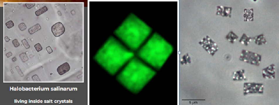
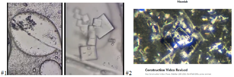
The shape-shifting morphology of  Haloarchaea “is comparable to that of bacteria and varies from rods, cocci, spirals to irregular shaped cells.”  Haloquadratum walsbyi (the squares shown center and right above) “is remarkable in that it forms an almost perfect quadrate flattened cell.” Other forms of the haloarchaea are shown below (A—D), and yes, some these individual varieties (if not all) change shape across multiple forms. Compare the images of H. salinarum shown in the post.

Image source, ‘Archaea –An Introduction’ https://www.sciencedirect.com/topics/immunology-and-microbiology/haloquadratum-walsbyi

“Halobacterium salinarum was originally grown in the laboratory from salted fish…” https://microbiologysociety.org/publication/past-issues/real-superheroes/article/the-immortal-halophilic-superhero-i-halobacterium-salinarum-i-a-long-lived-poly-extremophile.html

Want MA Gene?

(anagram of T-Mobile’s New Magenta)

In the Beginning of “Early Evolution on Earth — During the first half of Earth’s history, stretching over 2 billion years, dramatic and long-lasting evolutionary inventions occurred through processes that we are only beginning to understand (Fig. 1; Deamer, Reference Deamer2011; Knoll, Reference Knoll2015). They include prebiotic evolution and the development of cellularity, the foundation of the last universal common ancestor (LUCA) and evolution of the universal genetic code… [In] this paper, we discuss a speculative hypothesis for early evolution, called the ‘Purple Earth,’ which posits the rise of retinal pigment-based phototrophic life forms on Earth’s surface prior to anoxygenic and oxygenic photosynthesis.” https://www.cambridge.org/core/journals/international-journal-of-astrobiology/article/early-evolution-of-purple-retinal-pigments-on-earth-and-implications-for-exoplanet-biosignatures/63A1AD8AF544BEEF4C6D4A2D53130327 ;  The Purple Earth hypothesis is an astrobiological hypothesis that photosynthetic life forms of early Earth were based on the simpler molecule retinal rather than the more complex chlorophyll, making Earth appear purple rather than green.[1][2] An example of retinal-based organisms that exist today are the photosynthetic microbes collectively called Haloarchaea.[3] That time would date somewhere between 2.4 and 3.5 billion years ago, prior to the Great Oxygenation Event.[4] Many Haloarchaea contain the retinal protein, bacteriorhodopsin, in their purple membrane which carries out light-driven proton pumping, generating a proton-motive gradient across the cell membrane and driving ATP synthesis. The haloarchaeal purple membrane constitutes one of the simplest known bioenergetic systems for harvesting light energy. Retinal-containing purple membrane exhibits a single light absorption peak centered in the green-yellow energy-rich region of the visible spectrum, but allows transmission of red and blue light, resulting in a deep purple color… The Purple Earth hypothesis has great implications for the search for extraterrestrial life. …suggest[ing] that search methods should be expanded to planets reflecting blue and red light, since evolution of retinal-based photosynthesis is also probable, if not more likely than the evolution of chlorophyllic systems.” https://en.wikipedia.org/wiki/Purple_Earth_hypothesis

[2018] “Attempts to estimate when early life gained the ability to make the pigment [bacteriorhodopsin] – using a genetic mutation analysis technique called the “molecular clock” – have been hazy thus far, but the evidence suggests it was very soon after life emerged some 4 billion years ago. Similarly, researchers are unsure about the precise timeframe of chlorophyll’s emergence, but we do know that it happened at some point before 2.3 billion years ago – when, due to a combination of poorly understood factors, existing single-celled photosynthetic algae suddenly took over the ecosystem, causing what is called the Great Oxygenation Event. According to DasSarma and Schwieterman… ‘Evolution of anoxygenic photosynthesizers was followed by oxygenic photosynthesizing cyanobacteria and ultimately eukaryotic algae and plants,’ they wrote. ‘The development of eukaryotic algae and complex plants and their spread throughout the terrestrial environment allowed the evolution of land animals and ultimately intelligent life… This is exactly what got us thinking that the two pigments – retinal and chlorophyll – may have co-evolved,’ DasSarma added. To further investigate the “Purple Earth” hypothesis, he and Schwieterman propose future work focusing on natural communities of retinal-based phototrophs in diverse environments, as this may reveal unexpected roles and niches for this metabolism and inform its evolutionary origin.” https://www.iflscience.com/earths-first-life-forms-may-have-made-the-planet-purple-50225

Credit for ‘cornering’ and sequencing the archaeal LUCA ancestor gene was “the greatest scientist you’ve never heard of”Carl R.Woese (1928–2012), whom PBS/Nova called “The Man Who Rewrote the Tree of Life… having received a PhD in biophysics from Yale in 1953, Woese believed that there had to be a more objective, data-driven way to classify life… He arrived at the University of Illinois Urbana-Champaign as a microbiologist in the mid-1960s, shortly after James Watson and Francis Crick won the Nobel prize for their characterization of DNA’s double-helix form [in 1953]… In 1969, Woese wrote a letter to Crick, stating that: ‘…what I want to do is to determine primary structures for a number of genes in a very diverse group of organisms, on the hope that by deducing rather ancient ancestor sequences for these genes, one will eventually be in the position of being able to see features of the cell’s evolution.’… To create his evolutionary tree of life, then, Woese would need to choose a gene that was present in every known organism, one that was copied from generation to generation with a high degree of precision and mutated very slowly, so he would be able to track it over billions of years of evolution… Woese had several different rRNA molecules to choose from in the various subunits, which are classified based on their length. At around 120 nucleotides long, 5S rRNA wasn’t big enough to use to compare lots of different organisms. On the other end of the spectrum, 23S rRNA was more than 2300 nucleotides long, making it far too difficult for Woese to sequence using the technologies of the time. The Goldilocks molecule—long enough to allow for meaningful comparisons but not too long and difficult to sequence—was 16S rRNA in prokaryotes and its slightly longer eukaryotic equivalent, 18S rRNA. Woese decided to use these to create his quantitative tree of life. His choice was especially fortuitous, Eisen says, because of several factors inherent in 16S rRNA that Woese couldn’t have been aware of at the time, including its ability to measure evolutionary time on several different time scales. Certain parts of the 16S rRNA molecule mutate at different speeds. Changes to 16S rRNA are, on the whole, still extremely slow (humans share about 50% of their 16S rRNA sequence with the bacterium E. coli ), but one portion mutates much more slowly than the other. It’s as if the 16S rRNA clock has both an hour hand and a minute hand…”  https://www.pbs.org/wgbh/nova/article/carl-woese/

16S rRNA gene is universally encoded in all bacterial chromosomes.” https://journals.plos.org/plosone/article?id=10.1371/journal.pone.0202049

Carl Woese’s achievement for science was to find an undiscovered universe, a third New Kingdom of lifeforms whose genetic bounty suffuses other lifeforms like a parallel dimension: “In 1977, Carl Woese and George Fox published a brief paper in PNAS that established, for the first time, that the overall phylogenetic structure of the living world is tripartite… A fundamental breakthrough in biological science occurred in 1977, and most biologists did not notice… Several aspects of the paper by Woese and Fox (1) sparked skepticism. One was the arcane nature of the molecular data, which few could appreciate. The reliance on a single gene to trace major trends in evolution was an equally alien concept…” https://www.pnas.org/doi/10.1073/pnas.1109716109

But not for long.

Next-generation sequencing (NGS) is a technology for determining the sequence of DNA or RNA to study genetic variation…  Introduced for commercial use in 2005, this method …enabled… the interrogation of hundreds to thousands of genes at one time in multiple samples…quickly and cost-effectively… The speed, throughput, and accuracy of NGS has revolutionized genetic analysis…” https://www.thermofisher.com/us/en/home/life-science/sequencing/sequencing-learning-center/next-generation-sequencing-information/ngs-basics/what-is-next-generation-sequencing.html

[2019] “NGS methodology has been increasingly employed in the clarification of microbiota, especially in the gut. There are primarily two ways to detect bacterial DNA by using NGS methods. One approach is metagenome sequencing, which examines whole DNA of the microbial communities within a given sample without isolation or cultivation of individual species. The second approach is amplicon sequencing of the 16S ribosomal RNA (rRNA) gene (16S rRNA amplicon sequencing) since the 16S rRNA gene is universally encoded in all bacterial chromosomes.”https://journals.plos.org/plosone/article?id=10.1371/journal.pone.0202049

Here’s a brief gene sequencing tech timeline (from 1972 to the present) and the currently developing Third Generation (TGS) for single gene analysis https://frontlinegenomics.com/a-brief-history-of-next-generation-sequencing-ngs/

And when they find the genes of interest, they go fishing:

Fluorescence in situ hybridization (FISH) provides researchers with a way to visualize and map the genetic material in an individual’s cells, including specific genes or portions of genes… FISH is useful, for example, to help a researcher or clinician identify where a particular gene falls within an individual’s chromosomes. The first step is to prepare short sequences of single-stranded DNA that match a portion of the gene the researcher is looking for. These are called probes. The next step is to label these probes by attaching one of a number of colors of fluorescent dye.DNA is composed of two strands of complementary molecules that bind to each other like chemical magnets. Since the researchers’ probes are single-stranded, they are able to bind to the complementary strand of DNA, wherever it may reside on a person’s chromosomes. When a probe binds to a chromosome, its fluorescent tag provides a way for researchers to see its location.” (from the Human Genome Research Institute) https://www.genome.gov/about-genomics/fact-sheets/Fluorescence-In-Situ-Hybridization

SALT forming under microscopes

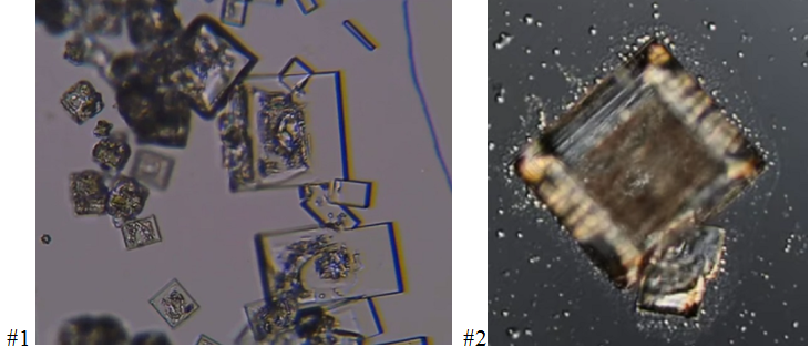
[1]https://www.youtube.com/watch?v=kGWBF5i7UzY

[2] https://www.youtube.com/watch?v=nC4rKdzqHr4

[3] https://www.youtube.com/watch?v=Vown1hyEyNw

[4](same as #1) https://www.youtube.com/watch?v=kGWBF5i7UzY

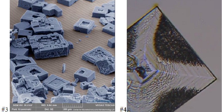
The Covid injection samples below look like salt

Is COVID technology messing with your salt?

Sodium, most of us know, is a key electrolyte in the proper function of our hearts, brains and cells. The body endeavors to continuously balance an optimal level of sodium by triggering its systems of fluid management. Most disruptions of sodium concentration in the blood lead to a state of hyponatremia which can be quickly fatal by seizures, heart and respiratory failure. I’ll leave you with some interactive Wikipedia links to make a quick study of problems and symptoms (mostly neurological and cardiac effects), and offer this glimpse of named conditions in sodium deficiency:

“Hyponatremia is a very common electrolyte disorder, especially in the elderly, and is associated with significant morbidity, mortality and disability. In particular, the consequences of acute hyponatremia on the brain may be severe, including permanent disability and death. Also chronic hyponatremia can affect the health status, causing attention deficit, gait instability, increased risk of falls and fractures, and osteoporosis. Furthermore, an overly rapid correction of hyponatremia can be associated with irreversible brain damage, which may be the result of the osmotic demyelination syndrome.” https://www.ncbi.nlm.nih.gov/pmc/articles/PMC4470176/

“Central pontine myelinolysis : “patients may present with the neurological signs and symptoms of hyponatraemic encephalopathy such as nausea and vomiting, confusion, headache and seizures. These symptoms may resolve with normalisation of the serum sodium concentration. …The classical clinical presentation is the progressive development of spastic quadriparesis, pseudobulbar palsy, and emotional lability (pseudobulbar affect), with other more variable neurological features associated with brainstem damage. These result from a rapid myelinolysis of the corticobulbar and corticospinal tracts in the brainstem…In about ten per cent of people with central pontine myelinolysis, extrapontine myelinolysis is also found. In these cases symptoms of Parkinson’s disease may be generated.” https://en.wikipedia.org/wiki/Central_pontine_myelinolysis ;

Hyponatremia or hyponatraemia is a low concentration of sodium in the blood…Symptoms can be absent, mild or severe. Mild symptoms include a decreased ability to think, headaches, nausea, and poor balance.[1][3] Severe symptoms include confusion, seizures, and coma; death can ensue.The causes of hyponatremia are typically classified by a person’s body fluid status into low volume, normal volume, or high volume.[4] Low volume hyponatremia can occur from diarrhea, vomiting, diuretics, and sweating.[4] Normal volume hyponatremia is divided into cases with dilute urine and concentrated urine.[4] Cases in which the urine is dilute include adrenal insufficiency, hypothyroidism, and drinking too much water or too much beer. Cases in which the urine is concentrated include syndrome of inappropriate antidiuretic hormone secretion (SIADH).[4] High volume hyponatremia can occur from heart failure, liver failure, and kidney failure.[4] Conditions that can lead to falsely low sodium measurements include high blood protein levels such as in multiple myeloma, high blood fat levels, and high blood sugar.” https://en.wikipedia.org/wiki/Hyponatremia

“…Causes of SIADH include conditions that dysregulate ADH secretion in the central nervous system, tumors that secrete ADH, drugs that increase ADH secretion, and many others. A list of common causes [includes]… Infections, Meningitis, encephalitis, brain abscess, rocky mountain spotted fever, AIDS, Perinatal asphyxia, Mass / bleed Trauma, subarachnoid hemorrhage, subdural hematoma, cavernous sinus thrombosis, Hydrocephalus, Guillain–Barré syndrome, Acute porphyria (acute intermittent porphyria, hereditary coproporphyria, variegate porphyria), Multiple system atrophy, Multiple sclerosis, Cancers …” https://en.wikipedia.org/wiki/Syndrome_of_inappropriate_antidiuretic_hormone_secretion https://en.wikipedia.org/wiki/Cerebral_salt-wasting_syndrome

“Hyponatremia would be common in patients who underwent radiotherapy for head and neck cancer” https://www.sciencedirect.com/science/article/pii/S2666555721000046    

–and that could apply to any irradiation of the head or body, even in times past by X-rays.

Intermittent hyponatremia occurs in a significant subset of patients with psychotic disorder as a consequence of a marked primary polydipsia and relatively minor and transient impairments in water excretion that appear linked to the underlying mental illness. Hyponatremia does occur in many other psychiatric patients as a consequence of medication-induced impairments in water excretion, which may or may not be aggravated by primary polydipsia. While effective preventative measures have been introduced, hyponatremia is frequently undetected in psychiatric patients, making the recognition and proper management an important, yet neglected, aspect of patient care.” https://link.springer.com/chapter/10.1007/978-1-4614-6645-1_9

“Polydipsia is excessive thirst or excess drinking.[1] The word derives from the Greek πολυδίψιος (poludípsios) “very thirsty”,[2] which is derived from πολύς (polús, “much, many”) + δίψα (dípsa, “thirst”). Polydipsia is a nonspecific symptom in various medical disorders. It also occurs as an abnormal behaviour in some non-human animals, such as in birds.” https://en.wikipedia.org/wiki/Polydipsia

…thirsty birds.


In 2007, with Next-Gen Sequencing in hand, Congress passed the NIH Reform Act:  “January 15, 2007 President Bush signed H.R. 6164 as P.L. 109-482, the National Institutes of Health Reform Act of 2006, affirming the importance of NIH and its vital role in advancing biomedical research…” https://www.nih.gov/about-nih/who-we-are/nih-reauthorization ; The following month, an organizational conference was held from February 9 to 11 called The Uncharted Microbial World and at the end of the year, on December 19, 2007, the NIH launched the Human Microbiome Project.

Feb 9-11, 2007- “The Uncharted Microbial World” https://www.ncbi.nlm.nih.gov/books/NBK562611/

“NIH Launches Human Microbiome Project” https://www.nih.gov/news-events/news-releases/nih-launches-human-microbiome-project

[Oct 2007] “The human microbiome project (HMP) reflects the fact that we are supraorganisms composed of human and microbial components. This international effort emanates from a confluence of ongoing technical and computational advances in the genome sciences… It is a global and interdisciplinary project that promises to break down the artificial barriers between medical and environmental microbiology… The human microbiome project (HMP) is an imposing yet logical conceptual and experimental extension of the human genome project… [that] will provide us with the parameters needed to design, implement and monitor strategies for intentionally manipulating our microbiota…” https://www.ncbi.nlm.nih.gov/pmc/articles/PMC3709439/

On the heels of the HMP came the formalization of the Earth Microbiome Project in 2010:

“Mapping the Microbiome of everything” –“The Earth Microbiome Project was founded in 2010 by Rob Knight, PhD, professor and director of the Center for Microbiome Innovation at UC San Diego; Jack Gilbert, PhD, professor and faculty director of The Microbiome Center at University of Chicago and group leader in Microbial Ecology at Argonne National Laboratory [partner with FermiLab synchrotron]; Rick Stevens, PhD, associate laboratory director at Argonne…and senior fellow at University of Chicago; and Janet Jansson, PhD, chief scientist for biology and laboratory fellow at Pacific Northwest National Laboratory [PNNL, the former Hanford Plutonium Works in Richland WA]…‘The potential applications for this database and the types of research questions we can now ask are almost limitless,’ Knight said.” https://www.uchicagomedicine.org/forefront/research-and-discoveries-articles/mapping-the-microbiome-of-everything 

 “Microbes en Masse…”[July 11, 2012] “Faeces, lizards, keyboards, faces — Rob Knight likes to sequence the microbes on anything and everything. Next, he plans to sequence Earth… Since 2010, Knight has been involved with the most ambitious effort yet to probe the microbiota: the Earth Microbiome Project, a collaborative effort to sequence… at least 200,000 environmental samples such as soils and water collected from around the world. He says that the project, which is led by Jack Gilbert…at the US Department of Energy’s Argonne National Laboratory in Illinois, will yield a master list of the proteins required to sustain microbial life across the planet;  about 500,000 reconstructed microbial genomes; and the makings of a global-scale model of microbial metabolism… Knight’s work certainly can make a splash: in 2010, the popular detective television programme CSI: Miami featured a technique developed by Knight and Noah Fierer, a microbial ecologist at Boulder, with which they matched individuals to the unique bacterial fingerprints that they left on computer keyboards11 (see ‘CSI: Microbes’). Knight’s team also attracted attention with as-yet unpublished work mapping the microbes that carpet the human face from forehead to lips…  Knight says that all his studies have a serious intent. The time-series data, for example, showed that there was vastly more variation within one person’s microbiota than expected… Janet Jansson, a microbial ecologist at the Lawrence Berkeley National Laboratory in Berkeley, California, [is] encouraging microbiologists worldwide to send in samples. They have already received some 60,000, ranging from deep-sea sediments from the Pacific Ocean to owl nests from Alaska… The plan, [Knight] says, is to collect a number of hypothesis-driven data sets, such as samples collected in pristine and disturbed areas… To that end, Knight continues to collect samples whenever he has the chance; later this month, for example, he will be scraping up microbial ‘mats’, which are among the most diverse communities known, from hypersaline waters off the Californian coast…  Knight has other ventures on the boil. In one project, he is exploring whether gut microbial communities can influence mental health by, for example, changing the signaling patterns between the gut and brain. “My family has schizophrenia on one side and bipolar disorder on the other,” he says, “which has led to my interest in trying to figure out whether we could potentially address those conditions.’ With Gordon and Lozupone, he is also part of a global network for the study of malnutrition and intestinal diseases, funded by the Bill & Melinda Gates Foundation in Seattle, Washington… Gilbert says that the research community faces a challenge in keeping up with Knight… “I think we’ll look back at him as a pioneer.” https://www.nature.com/articles/487156a#author-information

*

Going for the gut is nothing new. In 1896 a paper co-authored by Simon Flexner of the Rockefeller Institute begins, “The intestine, as will appear from these statements, is regarded as the portal of entry..[for] bacteria found in the inflamed peritoneum [and] also of some of those present in the pleura, upon the heart valves, and within the organs [of deceased on autopsy]…It has been found that these bacteria wander through the intestinal walls…where lesions of the intestinal mucosa exist… The majority of cases of acute pericarditis and acute endocarditis are found at the autopsy to be associated with pneumonia…” (https://collections.nlm.nih.gov/ext/dw/101317127/PDF/101317127.pdf )  In 1947, the eminent influenza researcher Frank Macfarlane Burnet recommended to his Australian government the use of intestinal agent bioweapons. (see the Influenza Special at the margin).

The great harvest for Big Science of the Microbiome expeditions, I contend, has to be the AMPs, another Rockefeller-sponsored endeavor based on the work of Rene Dubos, who discovered and isolated the first commercial antibiotics from Brevibacillus brevis in 1939. https://en.wikipedia.org/wiki/Ren%C3%A9_Dubos

“Antimicrobial peptides (AMPs): Ancient compounds that represent novel weapons in the fight against bacteria” –[June 2017]  Antimicrobial peptides (AMPS) are short peptide molecules produced by most living creatures. They help unicellular organisms to successfully compete for nutrients with other organisms sharing their biological niche, while AMPs form part of the immune system of multicellular creatures. Thus, these molecules represent biological weapons that have evolved over millions of years as a result of an escalating arms race for survival… hence, the appearance of resistance to these antimicrobials is rare…” https://www.sciencedirect.com/science/article/abs/pii/S000629521630301X?via%3Dihub

“Accurate and fast structure prediction of peptides of less [than] 40 amino acids in aqueous solution has many biological applications, but their conformations are pH- and salt concentration-dependent…” https://academic.oup.com/nar/advance-article/doi/10.1093/nar/gkad376/7160202?login=false

*

The engineering of micro-environments for salt and peptides appears to be a key feature of the biotech industry since the 1980s, contemporary with the major discoveries of opsin peptides from the haloarchaea and algae. Nowadays it’s become commonplace for the proteinaceous passengers in tiny salt vessels to be advantageously affected by the incorporation of artificial (non-natural, non-canonical) amino acids or nucleic acids in their design, giving them a survival edge in the Re-creation plan. If I’m reading it right, the ‘salt-box’ concept is to hold their activity in stasis through the ‘shipping’ stage by maintaining a salt environment until they can offload at their destination and reactivate. In some cases, possibly, for biosensor purposes or horizontal gene transfer, the salt package may be intended for a long-lasting voyage in your body. As Karen Kingston describes in Final Days, this technology is meant to function inside you but biologically independent of you, like a parallel dimension under intelligent control –a Second Life sharing your biological niche.

[2013]…” Here, we describe a strategy to boost salt resistance and serum stability of short antimicrobial peptides by adding the nonnatural bulky amino acid β-naphthylalanine to their termini…” https://pubmed.ncbi.nlm.nih.gov/23716061/

[2021] ”…In this work, we present a novel approach to include explicit water and salt in sequence-dependent coarse-grained (CG) models for proteins and RNA, enabling the study of biomolecular condensate formation in a salt-dependent manner… and to consider their key role in biomolecular condensate self-assembly.” https://pubs.aip.org/aip/jcp/article/155/12/125103/200724/Salt-dependent-phase-behavior-of-intrinsically

Rebooting life: engineering non-natural nucleic acids, proteins and metabolites in microorganisms” [2022]—“The surging demand of value-added products has steered the transition of laboratory microbes to microbial cell factories (MCFs) for facilitating production of large quantities of important native and non-native biomolecules. This shift has been possible through rewiring and optimizing different biosynthetic pathways in microbes by exercising frameworks of metabolic engineering and synthetic biology principles… to meet various therapeutic, biotechnological and industrial applications…” https://microbialcellfactories.biomedcentral.com/articles/10.1186/s12934-022-01828-y

According to Klaus Schwab (a name with a rough meaning of ‘flying talons’ or ‘attack from above’, born in Ravensburg), speaking at the 2023 World Government Summit: “... It’s just now that we move into the exponential phase… Whoever masters these technologies will be the master of the world.” –minute 30 of Final Days.

Watch the new documentary Final Days featuring Karen Kingston ( beginning at minute 24) where she highlights the practice of Directed Evolution: (min55) “In the very near future you’re going to be hearing this new term Directed Evolution… Directed Evolution is Transhumanism [and] if you are forcing nonhuman genetic mutations to our genome, you’re exterminating the human species.” https://www.stewpeters.com/video/2023/05/live-8-pm-et-final-days-worldwide-premiere/

Here’s an example of a typical research paper—

“Directed evolution of  rRNA improves translation kinetics and recombinant protein yield” [Sep2021] “Here, we evolve 16S ribosomal RNAs…from Escherichia coli, Pseudomonas aeruginosa, and Vibrio cholera towards enhanced protein synthesis rates…Directed evolution of ribosomal rRNA…towards unnatural bioactivities has highlighted plasticity in the cellular translation apparatus. Mutations to rRNAs or ribosomal proteins (r-proteins) can enhance translational fidelity, reduce translational kinetics, endow antibiotic resistance, affect ribosome assembly, and enhance the incorporation of noncanonical monomers or decode nonsense codons… A high-throughput methodology for directed evolution of rRNAs could …enable researcher-dictated biosynthetic capabilities… en route to unnatural bioactivities…” https://www.nature.com/articles/s41467-021-25852-5

_____________________

Revelations 3: 2 “Awake and strengthen what remains and is on the point of death… [Rev3:8-11]…Behold, I have set before you an open door which no one is able to shut; I know that you have but little power, and yet you have kept my word… I am coming soon; hold fast what you have so that no one may seize your crown.”

___________________________________________________________________

_______________________________________

END Notes, Part Four

The Shippers – The Allens and Rockefellers built their fortunes on shipping. Most of the high-tech mind control mentioned in The Minds of Men was funded under Navy contracts which operated the ONR, Office of Naval Research, whose establishment is attributed to Kuhn Loeb & Co banker Admiral Lewis L. Strauss. Strauss was Truman’s first choice of Atomic Energy Commission member in 1946. Not mentioned at all in The Minds of Men was Strauss’s hand-picked successor to the AEC, John A. McCone who became Allen Dulles’s replacement at CIA when JFK extended the offer, accepted November 29, 1961. McCone was an ultra-wealthy commercial shipping magnate at the time, integral to the blog post Atomic Oswald Three as a stakeholder in nuclear weapons and traffick. www.jenniferlake.wordpress.com/2018/01/29/atomic-oswald-three/

To this day, more than 30 years after his death (on Feb 14, 1991) McCone’s CIA-crafted biography remains classified and researchers on his trail claim they are tracked and censored on the net (see ‘education forum’ posts about McCone by John Simkin), and apparently he is still benefitting from the plausible deniability required with his CIA tenure. McCone thereafter continued as a CIA ‘consultant.’ Throughout his known career from involvement in the Six Companies (under Bechtel) that built the Hoover Dam, McCone was an early sponsor of nuclear development (making direct payments to Ernest Lawrence at UCBerkeley), member of Bohemian Grove and Knight of Malta. McCone is credited with establishing CIA’s Office of Science and Technology.

For a look at the Knights of Malta see David Whitehead’s serial documentary Cult of the Medics.  https://www.cultofthemedics.com/chapters.html Whitehead, incidentally, inserts pictures of himself ‘one-eyed’ and ‘hooded’ so take that with a grain of salt.

*

Korea came under multi-state foreign rule during World War II and appears to continue as a subject experimental population. Most of us know the Korean War narrative (June 1950 to July 1953) ushered in the demand for the Nevada Test Site and the mind control projects Bluebird and MKUltra.

Feb 2023— “South Korea’s world lowest fertility rate drops again… as of 2020, South Korea was the only country among the OECD members to have a rate below 1, giving it a shrinking population.” https://www.reuters.com/world/asia-pacific/south-koreas-world-lowest-fertility-rate-drops-again-2023-02-22/

*

SALT has enormous symbolic importance throughout time. The History Channel began its Big History series with salt as the foundation of civilizations and cause of all ‘roads’ made from animal trails to salt sources. The English place-name suffix ‘—wich’(or ‘wick’) stands for saltworks. Salt is variously symbolic of Earth, purity, protection, life and death. Holy Water is salted. The ‘symbolsage’ website offers this Norse myth among others: “According to Norse mythology, the gods were born of an ice-block, salty in nature, a process that took around four days to be completed. They were later brought to life as Adumbla, a cow, licked the salt and released them.” I’m reminded from this of the London Medical Society definition of ‘korona’ (Part 1 and above) https://symbolsage.com/salt-symbolism/and-meaning/

Multiple point mutations in microbes is called saltation evolution: “In biology, saltation (from Latin saltus ‘leap, jump’) is a sudden and large mutational change from one generation to the next, potentially causing single-step speciation” https://en.wikipedia.org/wiki/Saltation_(biology)

Is evolution always gradual or can it make leaps? We examine… the probability of multimutation leaps, that is, several mutations occurring simultaneously, within a single generation in 1 genome, and being fixed all together in the evolving population. The results indicate that… the probability of a multimutation leap is low, and accordingly the contribution of such leaps is minor at best. However, we show that, taking sign epistasis into account, leaps could become an important factor of evolution in cases of substantially elevated mutation rates, such as stress-induced mutagenesis in microbes. We hypothesize that stress-induced mutagenesis is an evolvable adaptive strategy.”  https://pubmed.ncbi.nlm.nih.gov/31570621/

*

The WEST NILE is the underworld burial ground of the dead.

In 1953  West Nile Virus was recovered from the delta by Stanford-trained Telford H. Work and Richard Moreland Taylor (Director of the Rockefeller Foundation International Health Division) under the auspices of U.S. Naval  Medical Research Unit Three in Cairo Egypt (est. 1942).

R.M. Taylor (papers): …”Diaries are missing for 1950-1952 and by 1953 Taylor was in Cairo, Egypt where NAMRU-3 is already established; there are no diaries covering the establishment of NAMRU-3 although there is a short description of its foundations in the 1956 diary. His diaries from 1953-1954 cover the bi-weekly activities of the lab…” https://findingaids.nlm.nih.gov/repositories/4/resources/736

The career and films of Telford Work https://medicineonscreen.nlm.nih.gov/2022/11/16/the-films-of-virologist-telford-work/ ; Here’s a more detailed biographical memoriam to Dr. Work https://archive.org/stream/cbarchive_103522_telfordhworkatribute1996/JAMCA_V12_N3_P385-395_djvu.txt

Below is a line graph of West Nile Virus.


The “public-private”department of government organizing the comprehensive data collection designated for vaccine passports is called the National Interoperability Collaborative, https://nic-us.org and here’s its logo.

*The NIC is managed under the auspices of the Stewards of Change Institute, SOCI, that happens to reverse to twilight-language-like ICOS, adding possible meaning to “icosahedron” and the ICOS immune complex (standing for “Inducible CO-Stimulator” signal pathway. More on this in a later post)

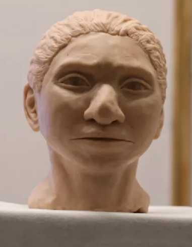
Another “MA gene” candidate comes not from bacteria but from a human Denisovan:

MA-1 gene:

[2013]“…Here we sequence the draft genome of an approximately 24,000-year-old individual (MA-1), from Mal’ta in south-central Siberia9, to an average depth of 1×. To our knowledge this is the oldest anatomically modern human genome reported to date. The MA-1 mitochondrial genome belongs to haplogroup U, which has also been found at high frequency among Upper Palaeolithic and Mesolithic European hunter-gatherers10,11,12, and the Y chromosome of MA-1 is basal to modern-day western Eurasians and near the root of most Native American lineages5. Similarly, we find autosomal evidence that MA-1 is basal to modern-day western Eurasians and genetically closely related to modern-day Native Americans, with no close affinity to east Asians. This suggests that populations related to contemporary western Eurasians had a more north-easterly distribution 24,000 years ago than commonly thought…” https://www.nature.com/articles/nature12736/

First to do it, an Israeli team established at Hebrew University produced “The artistic rendering of the head and face of a 13-year-old girl from the prehistoric… Denisovans, based on technology developed by Hebrew University professor Liran Carmel and his team. The bust was revealed at a news conference in Jerusalem on Sept. 19, 2019 [9-19-19]” https://www.livescience.com/denisovans-extinct-human-relative ; Team member David Gokhman, currently at the Weizmann Institute, specializes in cellular hybrids of human-apes with a focus on brain and neuronal structures. https://www.weizmann.ac.il/WeizmannCompass/sections/new-scientists/dr-david-gokhman; (example) “Here we utilized human-chimpanzee hybrid induced pluripotent stem cells to study gene expression separating these species…and to control for nongenetic confounding factors. We differentiated these cells into cranial neural crest cells, the primary cell type giving rise to the face….” https://pubmed.ncbi.nlm.nih.gov/33731941/

Remember the unexplained “Chromosome 8” marker gene target from the Covid PCR tests? A particular distinction of chromosome 8 separates humans from other primates.

*

Build Back a Better (?) Brain:

How to design and build abnormal brains using radiation during
development. In Disorders of the Developing NervousSystem,
Fields, W.S. and Desmond, M.M., eds..  Springfield, Ill:Thomas,
1961.  Also available as US Atomic Energy Comm TID6159:1-28, Jun
60; 
  pp. 60-97.##”

–Notice the year date 1960 while McCone was AEC chairman and AEC security was peopled with ONI (Office of Naval Intelligence). I singled out this citation above for the ‘polio forever’ blog from a bibliographic source in GWU’s “national archive” used by President Clinton’s Advisory Commission on Human Radiation Experiments (ACHRE) https://nsarchive2.gwu.edu/radiation/dir/mstreet/research/pubs/titles.txt The two researchers responsible for this 1960 study are Samuel P. Hicks and Constance J. D’Amato, and while I can’t find the text of “How to…build abnormal brains”, a search on either Hicks or D’Amato yields similar material.

Examples for Hicks: Research works by Samuel P. Hicks https://www.researchgate.net/scientific-contributions/Samuel-P-Hicks-2061151130 ; Obit from UMichigan –Naval officer at Strong Memorial Hosp in Rochester NY during WWII (infamous for plutonium feeding), US Navy pathologist (1946-47), Naval Medical Center, etc. https://record.umich.edu/articles/obituary-samuel-p-hicks/ ; 1968 brain study under AEC contract https://onlinelibrary.wiley.com/doi/10.1002/ar.1091600311

…more END NOTES to Part Four and a forthcoming epilogue of the Crow are in progress…..

*

*

*

May 27, 2021

Born Old

*

Can ‘normal’ children be born with ‘old’ diseases? Longevity research which lumps Alzheimers and atherosclerosis together with inflammatory asthma, for example, suggests they can by focusing on the role of NAD-dependent sirtuins: “[Sirtuins] mute the chronic, overactive inflammation that drives diseases such as atherosclerosis, metabolic disorders, ulcerative colitis, arthritis, and asthma.” –p24, Lifespan, by David Sinclair, 2019

I once overheard two new mothers pushing their baby strollers past my house and chatting loudly during their early-morning workout: “But the doctor said he was born with asthma!” said one. “Not only that but, did you know…” said the other, and the conversation trailed off with them as they moved. My mind fixed on the words ‘born with asthma’. This was 10 years ago and I still haven’t found a mainstream citation to support the claim, though admittedly not looking hard for it. Asthma, they say, is caused by viruses, and babies indeed are born with those —  apparently  born with a variety of tobacco-type viruses (tobamovirus) in their guts which increase in the first year of life to as much as 50% of viral content in the gut biome as they near age 2.

Tobamoviruses come from plants, it is assumed. Vectors known today include any number of members from all the Life kingdoms.

Asthma, at face value:

Asthma in Children: Risk Factors, Diagnosis, Management

https://my.clevelandclinic.org/health/diseases/6776-asthma-in-children

Asthma is the leading cause of chronic (long-term) illness in children. It affects more than 7 million children in the United States. For unknown reasons, the rate is steadily increasing. Asthma can begin at any age, but most children who have it have their first symptom by age 5.

*

Childhood asthma – Symptoms and causes – Mayo Clinic

https://www.mayoclinic.org/diseases-conditions/childhood-asthma/symptoms-causes/syc-20351507

In childhood asthma, the lungs and airways become easily inflamed when exposed to certain triggers, such as inhaling pollen or catching a cold or other respiratory infection. Childhood asthma can cause bothersome daily symptoms that interfere with play, sports, school and sleep.

*

Asthma in Infants | AAFA.org

https://www.aafa.org/asthma-in-infants/

Some preschool children get viral infections often. At least half of children with asthma show some sign of it before the age of 5. Viruses are the most common cause of acute asthma episodes in infants 6 months old or younger. How Is Asthma in Infants and Toddlers Different Than Adult Asthma?

*

Viral Infections and Associated Factors That Promote Acute …

https://pubmed.ncbi.nlm.nih.gov/29178673

Early childhood infections with well-known or emerging viruses can lay the pathophysiologic framework for asthma development and exacerbation later in life, which may be due partly to alteration of the airway microbiome. Once asthma is established, acute exacerbations are usually associated with infections with respiratory viruses, such as rhinoviruses (RVs)…

*****

Rhinoviruses are identical to polioviruses, which in turn are identical to certain plant viruses –read on to citation “Can Plant Viruses Cross the Kingdom Border and Be Pathogenic to Humans.”

*****

[2009] Bugs and asthma: a different disease?

“The prevalence of asthma has dramatically increased in recent decades. Exacerbations of asthma are a large contributor to asthma-related costs, and are primarily caused by viral and atypical bacterial infections. Rhinoviruses (RVs) are the most common viruses detected after an asthma exacerbation. RVs, respiratory syncytial virus (RSV), and human metapneumovirus (hMPV) viral infections early in life can induce wheezing and are associated with the development of asthma later in life. Atypical bacterial infections from Mycoplasma pneumoniae and Chlamydia pneumoniae have also been linked to chronic asthma and potential asthma exacerbations. In this article, we will discuss recent developments in viral infections, specifically RV, RSV, and hMPV, and atypical bacterial infections as causes of asthma…” https://pubmed.ncbi.nlm.nih.gov/19387028/

*******

Life in the Virosphere

Longevity molecules are being discovered in “Plants experiencing stress”—from Lifespan: “[R]esveratrol is produced in greater quantities by grapes and other plants experiencing stress. We also knew that many other health-promoting molecules, and chemical derivatives of them, are produced in abundance by stressed plants; we get resveratrol from grapes; aspirin from willow bark, metformin from lilacs… Plants have survival circuits too, and we think we might have evolved to sense the chemicals they produce in times of stress as an early-warning system, of sorts, to alert our bodies to hunker down [in ‘protection’ mode] as well. What this means, if it’s true, is that when we search for new drugs [or new microbes] from the natural world we should be searching the stressed-out ones: in stressed plants, in stressed fungi, and even in the stressed microbiome populations in our guts. The theory is also relevant to the foods we eat; plants that are stressed have higher concentrations of xenohormetic molecules…” –p131, ibid.

*

Hormesis is a characteristic of many biological processes…[in] response to exposure to increasing amounts of a substance or condition…[and is]  generally a favorable biological response to low exposures to toxins and other stressors. The term hormesis comes from Greek hórmēsis “rapid motion, eagerness”, itself from ancient Greek hormáein “to set in motion, impel, urge on”… https://en.wikipedia.org/wiki/Hormesis

—in other words, accelerating stress.

“This review compiles information on how hormesis in plants can be used to achieve new production levels… [by application of pesticides, such as glyphosate, where]..the plants benefit from a low dose whereas are distressed by a high dose which leads to an irreversible and negative outcomes on the health and functions of plants” https://www.sciencedirect.com/science/article/pii/S0147651320310642

Directing hormetic activity by inducing stress ‘toxins’ with chemicals and radiation particularly,  is a controversial practice, not to mention other “trauma-based hormesis.” Largely from insights on what is posted here, hormetic “stress chemicals” produced in response to change are another addition to the category of ‘virus’ .

*************

The dominant ‘birth’ virus identified in the Nature article below (Aug.2020) and drawn from a small study in 2016, showed Pepper Mild Mottle Virus (PMMoV) as the most abundant ‘reconstructed’ genome, but the classic ‘type species,’ Tobacco Mosaic Virus (TMV) was also found in these infants, though their parents neither smoked nor handled tobacco. By size and shape, PMMoV and TMV are identical. TMV, in rod form, can be extracted from many hundreds of common food plants. There are also disc, ring, and spherical virions of TMV, but that subject will be explored in the next installment (#4) of Planting Viruses—to some extent, already posted in #3.

[2009]…” Typical tobamovirus-like particles (rigid rods ≈300 nm long) were observed in crude plant extracts. According to literature, at least six tobamoviruses infect peppers: Paprika mild mottle virus (PaMMV); Pepper mild mottle virus (PMMoV); Ribgrass mosaic virus (RMV); Tobacco mild green mosaic virus (TMGMV); Tobacco mosaic virus (TMV); and Tomato mosaic virus (ToMV)” …  https://pubmed.ncbi.nlm.nih.gov/30764375/

New ‘pepper’ tobamoviruses have recently been discovered [2008] and this citation is relevant to my grocery shopping—the packaged peppers on my area’s food shelves are imports from Israel.

[2008] “The biological, serological, and molecular characteristics of a newly isolated L4 resistance-breaking isolate of Pepper mild mottle virus (PMMoV) were studied. The new pathotype of PMMoV is closely related to the Israeli pathotypes P1,2 and P1,2,3 of the virus; however, the mosaic symptoms caused by this new pathotype on pepper plants with an L4 genotype were more severe than the mild mosaic symptoms caused by other common pathotypes…”  https://www.apsnet.org/publications/plantdisease/2008/July/Pages/92_7_1033.aspx

[2010] Pepper Mild Mottle Virus, a Plant Virus Associated with Specific Immune Responses, Fever, Abdominal Pains, and Pruritus in Humans

“Recently, metagenomic studies have identified viable Pepper mild mottle virus (PMMoV), a plant virus, in the stool of healthy subjects. However, its source and role as pathogen have not been determined…” https://journals.plos.org/plosone/article?id=10.1371/journal.pone.0010041

**************

[2015]

Can Plant Viruses Cross the Kingdom Border and Be Pathogenic to Humans?

…” Accordingly, plant viruses are not considered to present potential pathogenicity to humans and other vertebrates. Notwithstanding these beliefs, there are many examples where phytoviruses circulate and propagate in insect vectors. Several issues are raised here that question if plant viruses might further cross the kingdom barrier to cause diseases in humans. Indeed, there is close relatedness between some plant and animal viruses, and almost identical gene repertoires. Moreover, plant viruses can be detected in non-human mammals and humans samples, and there are evidence of immune responses to plant viruses in invertebrates, non-human vertebrates and humans, and of the entry of plant viruses or their genomes into non-human mammal cells and bodies after experimental exposure. Overall, the question raised here is unresolved…It is currently considered that phytoviruses only infect plants and therefore, plant viruses cannot cause disease in humans. In the present review, several issues are raised that challenge this paradigm.…”We describe here that some plant and animal viruses are closely related; humans are considerably exposed to plant viruses; plant viruses can enter mammalian cells and bodies and be naturally present in mammals, including humans; and this presence may be non-neutral; plant viruses may trigger events in mammalian cells and elicit immune responses in invertebrates, non-human vertebrates and humans, and was recently associated with clinical symptoms.

…”The distribution of plant and animal viruses within the currently defined taxons indicates that some plant and animal viruses belong to same viral families (Table 1)…  This is true for family Reoviridae, which includes rotavirus, a major cause of gastroenteritis in humans [36], family Rhabdoviridae, which notably includes rabies virus [37], and family Bunyaviridae, which encompasses human pathogens such as Hantaan virus and Toscana virus [38,39,40]. Within the family Rhabdoviridae, genomic organization indicates that some plant viruses of genera Nucleorhabdovirus and Cytorhabdovirus differ from animal viruses of genera VesiculovirusLyssavirusNovirhabdovirus and Ephemerovirus only by the presence of an additional gene coding for a movement protein, a protein essential for the systemic spread of the virus in the host plant [17,18,37,41]….

…”In addition, Cowpea mosaic virus (CPMV), which is a member of family Secoviridae, shares structural features and genetic organization with animal picornaviruses such as polioviruses, coxsackie viruses and Theiler’s murine encephalomyelitis virus (TMEV), which supports the hypothesis that these plant and animal viruses might derive from a common ancestor [43,44]. In addition, pararetroviruses include plant viruses from the family Caulimoviridae, which consists of the only plant viruses with a double-stranded (ds) DNA genome, as well as animal viruses that belong to the family Hepadnaviridae, which includes hepatitis B virus [45]. Similarly, hepatitis E virus, a leading cause of acute hepatitis in humans, was grouped with Beet necrotic yellow vein virus, a plant virus that belongs to the genus Benyvirus, based on phylogeny of RNA polymerases [46]. Recently, endogenous viral elements (EVEs) from plant RNA viruses were detected in 14 insect genomes [47]. Interestingly, EVE sequences very close to those of phytoviruses of the family Virgaviridae were detected in the genome of a mosquito, Aedes aegypti; a fly, Drosophila rhopaloa; and a bee, Bombyx terrestris. The EVE detected in Aedes aegypti was similar to the tobamovirus genome, which is very surprising as no insect vector is known for tobamoviruses…”

https://www.mdpi.com/1999-4915/7/4/2074/htm

*************

NATURE magazine, August 2020

 “In this work, we report that plant viruses can also be found frequently in the gut and oropharynx of children during their first year of life, even when they are exclusively breast-fed…

“In 100% of the fecal samples and 65% of the oropharynx samples at least one plant virus was identified. Tobamoviruses in the Virgaviridae family were by far the most frequently detected, with tropical soda apple mosaic virus, pepper mild mottle virus, and opuntia tobamovirus 2 being the most common species. Seventeen complete virus genomes could be assembled, and phylogenetic analyses showed a large diversity of virus strains circulating in the population. These results suggest that children are continuously exposed to an extensive and highly diverse collection of tobamoviruses…

“Metagenomic studies describing the composition of eukaryotic viruses in the gastrointestinal and respiratory tracts of children are limited, and only a few of them have included children under 1 year of age5,6,7,8,9,10,11. In addition, most of the reported studies have characterized the virome in diseased children8,9,10,11 and just a few have studied community-based healthy children5,6,7. Therefore, we aimed to determine the diversity and dynamic of the oropharyngeal and gastrointestinal eukaryotic viromes in [healthy] children under 1 year of age in a semi-rural population in Mexico, using next generation sequencing. In addition to human viruses, we found that plant viruses were commonly present in the gut and the oropharynx of children during their first year of life and, surprisingly, they were found as early as 2-weeks after birth in exclusively breast-fed infants. Tobamoviruses, in the Virgaviridae family, were the most abundant, and were present in most of the samples analyzed. Of interest, antibodies to plant viruses have been found in animals, including humans3, and it has also been shown that cowpea mosaic virus can disseminate systemically when orally administered to mice12. Whether the common presence of these viruses at an early age has an effect in the infant’s immune system and maturation of the gut remains to be investigated…

…”Some studies have detected the presence of Virgaviridae sequences in fecal samples of children with particular pathologies. Thus, in a study carried out in the Hunan province of China, 238 fecal samples were collected from pediatric patients (0–12.4 years of age) who were clinically diagnosed with mild or severe hand-foot-and-mouth disease10. In pools of samples from severe cases, 94% of the total viral reads were classified in the family Virgaviridae…

“It is now clear that the bacterial communities in the gastrointestinal tract play a critical role on different human developmental pathways early in life22. In this regard, it is of great interest the considerable exposure that infants can have to plant viruses during the first year of life, as shown by the high frequency of detection of tobamoviruses in both the oropharynx (65%) and fecal (100%) samples starting at least 2 weeks after birth. This raises the question of whether these viruses could have an impact in the maturation of the infant’s immune system or in their healthy development, directly or through interactions with other components of the microbiota, as has been observed for norovirus [Norwalk virus] in a mouse model23. This possibility needs to be investigated.”

(snip) https://www.nature.com/articles/s41598-020-70684-w

*

This Nature study excerpted above, like all others purporting to study humans, makes Zero mention of the vaccination status of study subjects. Given its recent date though, it can and should be assumed that participants were vaccinated according to WHO/CDC recommendations. At the moment, Mexico exempts children under 18 from COVID vaccines unlike the U.S. which is now recommending CoVax for 6-month-old babies and pregnant women.

Routine shots for babies at birth in Mexico, now for longstanding, includes BCG (tuberculosis) and HepB. At 2-months they get Rotarix (rotavirus), anti-pneumococcal, and a whopping ‘pentavalent’ vaccine against H.influenzae, pertussis, diphtheria, tentanus and polio. At six months of age they start regular vaccines for influenza and polio (Sabin OPV and Salk IPV). At 12-months adding HepA, varicella (chicken pox) and Triple SRP (measles, mumps, rubella) –and after that, boosters as they grow, HPV at age eleven and so on. https://en.wikipedia.org/wiki/Vaccination_in_Mexico

Most of these vaccines would also have been given to the mothers when they were young and since 2012, pregnant mothers receive an additional Tdap. The ‘Nature’ study picks up its pregnant volunteers in the third trimester—after, presumably, getting their Tdap.

·  Pregnancy and Vaccination: Why Maternal Vaccines Are …

http://www.cdc.gov › vaccines › pregnancy

Tdap Vaccine Tdap vaccine is recommended during every pregnancy, between 27 and 36 weeks gestation (preferably earlier in this period). When given during pregnancy, Tdap vaccine boosts antibodies in the mother, which are transferred to her developing baby. Early third trimester administration optimizes neonatal antibody levels.”

******

Before I launch my ‘stress chemical’ explanation for the presence of tobamoviruses in newborns, I’d like to share this complex description of the mRNA CoV vaccines from an interview between Joseph Mercola and Stephanie Seneff.  Touching on a lot of points of virus interaction, this interview also has a beautiful illustration of how our bodies integrate RNA, reverse transcribe it to DNA and pass the genetic ‘signals’ (of more RNA and proteins) on to other cells, including sperm and egg cells.

https://articles.mercola.com/sites/articles/archive/2021/05/23/stephanie-seneff-covid-vaccine.aspx

*

Minute 29—

Mercola: “You found that there is a lot of [naturally-existing] reverse transcriptase in our [cellular] DNA…and this causes our body to turn RNA back into DNA…

Seneff: “…we actually have plenty of reverse transcriptase in our own cells… and if these LINEs and SINEs are able to [convert] RNA back to DNA and to put that DNA back into the genome… and those [LINEs, “sometimes called ‘junkDNA’”] …surprisingly is 10% of our DNA… they’re really weird—they fold DNA and stick it in backwards and…do crazy stuff [that’s] so fascinating…  And when people have Alzheimers…[for example] they get multiple copies of that amyloid beta protein…[as] extra genome, with variations in those copies [which] they [made] through RNA…  It’s an evolution, and the mechanism from which we evolve, probably… So, we’re taking that RNA, mutating it –because RNA mutates much more easily than DNA does—and then [transcribing] it back into DNA and sticking it back into the genome… This is a known process associated with cancer and all these neurological diseases…

“And when you have [glyphosate, i.e.] with the body so sick [it’s] trying to mutate its way around those problems… It’s a process we use to try and deal with toxic chemicals in [our] environment…

Mercola: “…So,[mutations] can be transferred down genetically…like editing…

Seneff: “yes, and I was blown away to learn about the sperm… There’s a complete story in it that sperm can take… messenger RNA, external RNA and that can be from a virus or a vaccine, taking it in and converting it to DNA and then producing what’s called plasmids. So the sperm does this: whatever mRNA they come up with…,they make [into] plasmids…which is converted to DNA and put into these little pellets –and all the sperm [not just the ‘lucky’ one] in this [fertilizing] environment [around the egg]…are basically handing over to the egg all these plasmids with nuggets of RNA/DNA in them that they [picked up]… The egg takes it up and puts them into all the cells as it grows and… by the time the child is born, it’s got all these plasmids in there with…code to make [vaccine/viral] proteins… And now that child is not going to have any antibodies to that protein –its going to [detect only] human protein. It’s immune system is going to be trained that this is a natural protein and doesn’t need antibodies to it. So if that child gets exposed to [the corresponding] disease it’s immune system won’t react…  and they’ll be able to carry that virus for their entire lives and pass it on down to their children…

…”Now, there is [a] disease that cows were getting—a diarrheal infection that was a problem—and what would happen is a baby calf would be born with that virus protein belonging to that genome and the calf would carry that virus and spread it to all the cows, so [farmers] became aware that [they] had all these ‘killer’ calves being born and the adult cows were getting sick with the infection that the calves were carrying… So, I think, this can be done with [humans]…

Mercola: …”basically [creating] superspreaders…”

Seneff: “Yes, and they killed these calves…when they found them…because they couldn’t afford to infect an entire herd…with this virus…  So, a virus goes through this process; at first it’s new to the population and causes…disease and …eventually there’s, I think, something in a virus that’s needed , potentially, to get into [the body] system to help cope [with toxins]. 

–end transcript at min.37

*************************************************************************************

You Are What You EAT –and what eats you!

In light of the ‘healthy’ TMV-like content of baby guts, scrupulous consideration should be given to the current manipulation of tobamoviruses in plants and the discovery of vectoring these “plant” viruses by mosquitos!

Firstly, there’s the “flying syringe” funded in 2010:

·  ‘Flying Syringe’ Mosquitoes Get Bill Gates Funding

http://www.bibliotecapleyades.net/ciencia/ciencia…

“The Bill and Melinda Gates Foundation awarded 100,000 dollars each on Wednesday to scientists in 22 countries including funding for a Japanese proposal to turn mosquitoes into “flying syringes” delivering vaccines. The charitable foundation created by the founder of software giant Microsoft said in a statement that the grants were designed to, “explore bold and largely unproven ways to improve global health.”

*

—And then consider decades of experimentation on GMO food and crops with a newer link below to “foreign protein expression” obtained in tobacco: This citation appears to spin out of the same academic network as ’flying syringes’:

[Title] “Improvement of the transient expression system for production of recombinant protein in plants”… [text]”Transgenic plants are generally used to obtain recombinant proteins or identify the localization of proteins…The replication system of plant viruses results in high-level expression of foreign proteins within a few days..[and] can be easily amplified…[such as the] tobamovirus (TMV)-based deconstructed viral system [called] magnIcon [which] has been extensively engineered to achieve high levels of recombinant protein…[demonstrated by the vaccine] ZMapp for Ebolavirus infections…used during a recent outbreak… [Africa, 2014]” https://www.nature.com/articles/s41598-018-23024-y

*

And then there’s evidence of tobacco use 12,000 years ago — do you think– as food?

Oldest Known Evidence of Tobacco Use in North America Found in Ice Age Hunting Camp

…. “the most interesting part of the discovery is that there is no direct evidence that people used tobacco past 3,000 years ago, but this research proved its use more than 12,000 years ago.”

https://www.ancient-origins.net/news-history-archaeology/oldest-known-evidence-tobacco-use-north-america-found-ice-age-hunting-camp-020922

******

“You are what you eat: Sequence analysis reveals how plant microRNAs may regulate the human genome”

March 2019

Abstract

“Background: Nutrigenomic has revolutionized our understanding of nutrition. As plants make up a noticeable part of our diet, in the present study we chose microRNAs of edible plants and investigated if they can perfectly match human genes, indicating potential regulatory functionalities… Results: Four common plant miRNAs were identified to match perfectly with 22 human transcripts. The identified target genes were involved in a broad range of body functions, from muscle contraction to tumor suppression. We could also indicate some connections between these findings and folk herbology and botanical medicine…”

https://pubmed.ncbi.nlm.nih.gov/30708219/

******

“Plant miRNAs found in human circulating system provide evidences of cross kingdom RNAi”

March 2017

Background: Emerging evidence indicates that plant miRNAs can present within human circulating system through dietary intake and regulate human gene expression…  Results: In this study, we identified abundant plant miRNAs sequences from 410 human plasma small RNA sequencing data sets. One particular plant miRNA miR2910, conserved in fruits and vegetables, was found to present in high relative amount in the plasma samples… Conclusions: Through analysis of public available plasma small RNA sequencing data, we found the supporting evidence for the plant miRNAs cross kingdom RNAi within human circulating system.”  https://pubmed.ncbi.nlm.nih.gov/28361700/


**************************************************

The Long and Short of RNA

On the frontier of biological complexity is an ‘RNA world’ that prior to the 1980s, research scientists knew little about. They have since learned that most of the genetic material we carry is non-coding, meaning there are no specific proteins associated with it. This is our extraordinary and dynamic epigenome that directs cell functions, behavior, and identity.  Professor John Mattick says in his presentation, The New World of RNA Biology, that “most of the genes in humans do not code for proteins”. He compares the known human “coding regions” with those of nematodes (hookworms, for example )as being numerically equivalent, “which means that [we have] more regulatory genes than genes –which is impossible [it was thought]…so, there’s another conversation about how eukaryotes solve this problem which is through microRNAs…and modularity.”  Here’s his wonderful [one hour] ‘classroom’ video  https://www.youtube.com/watch?v=WdltOXdwo4g

https://en.wikipedia.org/wiki/John_Mattick

RNA interference –RNAi

“RNAi encompasses an array of ancient and sophisticated cellular mechanisms that regulate a variety of biological functions. Argonaute proteins bind many naturally occurring small RNAs to defend against transposable elements, maintain chromosome structure and stability, and regulate developmental timing and differentiation. For example, microRNAs [miRNA] represent a natural form of developmentally-important siRNAs [small interfering RNA]…  [O]ne microRNA [can] regulate hundreds of genes. Humans make more than 500 distinct microRNAs, and the inappropriate production of specific microRNAs has been linked to several diseases…” https://www.umassmed.edu/rti/biology/how-rnai-works/

RNAi, 10 minute video explanation by Maria Portela https://www.youtube.com/watch?v=C-pTiFkg1aU

A basic understanding of ‘RNA world’ is enough to follow the logic of what I’ll call “virus world,” as the lexicon of viruses has indeed absorbed the ‘transcript’ fragments from noncoding RNA (i.e. retrotransposons=endogenous viruses=’jumping genes’, etc.) and the endless variety of enzymes and proteins that are made for our adapting and evolving survival.

According to Dr. Mattick, the structural RNAs which build and shape our genes are those most familiar and studied by science: “There are studies going back to the 1960s showing that there are very stable chromatin-associated RNAs… RNA is the third component of chromatin and it’s nuclease resistant”, so it remains stable, resistant to enzymatic degradation—and those are qualities that make Tobacco Mosaic Virus so very interesting. TMV, in fact, looks like a chromatin stack. More than that, it has an observable relationship with the presence of human beings all over the world and deep into antiquity. TMV has more ‘mysteries’ than just its own genetics to reveal.

chromatin coiled-coil structure

************************************************************************************

Ebola, if you’re wondering, looks like TMV. I’ll have more to say (and show) about it in a future ‘Planting Viruses’.  A particular TMV mystery is the “coat protein” homologue amino sequence that matches the human/mitochondria enzyme ‘TOMM40L’ found on chromosome 1.  (ref. https://pubmed.ncbi.nlm.nih.gov/23573274/) The human/plant construct TOMM40L is known as a “translocase”:

“Translocase is a general term for a protein that assists in moving another molecule, usually across a cell membrane. These enzymes catalyze the movement of ions or molecules across membranes or their separation within membranes… It is also a historical term for the protein now called elongation factor G, due to its function in moving the transfer RNA (tRNA) and messenger RNA (mRNA) through the ribosome… Several of these [translocases] involve the hydrolysis of ATP and had been previously classified as ATPases (EC 3.6.3.-), although the hydrolytic reaction is not their primary function…

[Translocase Example #3 (of 3, at the bottom of the page): “Translocase of outer mitochondrial membrane 40 (TOMM40), a protein encoded by the TOMM40 gene, [is an enzyme] whose alleles differentially impact the risk for Alzheimer’s diseasehttps://en.wikipedia.org/wiki/Translocase Alzheimer ‘genes’ also encode on Chromosome 1.

And this is just one example, due probably to the extra attention of medical science on Alzheimers. In 2008 I read a special edition feature in the Los Angeles Times reporting that Americans then in early middle age could expect a 100% rate of Alzheimers.

*

Chromosome 1 is the designation for the largest human chromosome… It was the last completed chromosome, sequenced two decades after the beginning of the Human Genome Project… There are 890 known diseases related to this chromosome” –including porphyria and the single-point mutation disease of rapid premature aging called Hutchinson-Gilford Progeria. https://en.wikipedia.org/wiki/Chromosome_1

So far, however, there is only one claim of a completely sequenced chromosome, and it’s not chromosome 1. This past April it was widely published that chromosome 8 is the first –the additional entity sought by the COVID PCR tests.

  Complete Chromosome 8 Sequence Reveals New Genes and Disease …

https://phys.org/news/2021-04-chromosome-sequence-reveals-genes-disease.html

************************************************************************************

Look again at chromatin structure as it’s bound into a chromosome. Structure, of itself, is the manifestation of a chemical ‘story’.

*

Building with RNA

Architectural RNA in chromatin organization – “RNA plays a well-established architectural role in the formation of membraneless interchromatin nuclear bodies. However, a less well-known role of RNA is in organizing chromatin, whereby specific RNAs have been found to recruit chromatin modifier proteins… Studies… suggest that RNA plays a major direct architectural role in chromatin organization…” https://pubmed.ncbi.nlm.nih.gov/32897323/

*

“Mammalian transcriptome analyses elucidated the presence of thousands of unannotated long noncoding RNAs (lncRNAs) with distinct transcriptional units. Molecular characterization and functional classification of these lncRNAs are important challenges in the next decade. A subset of these lncRNAs is the core of nuclear bodies, which are the sites of the biogenesis, maturation, storage, and sequestration of specific RNAs, proteins, and ribonucleoprotein complexes. Here, we define a class of lncRNAs termed architectural RNAs (arcRNAs) that function as the essential scaffold or platform of nuclear bodies. Presently, five lncRNAs from mammals, insects, and yeast are classified as arcRNAs. These arcRNAs are temporarily upregulated upon specific cellular stresses, in developmental stages, or in various disease conditions, and sequestrate specific regulatory proteins, thereby changing gene expression patterns.” https://pubmed.ncbi.nlm.nih.gov/26021608/

*

“It is now evident that transcriptional gene regulation usually requires the re-organization of chromatin architecture. Increasing evidence suggested various kinds of RNAs are involved in this process. Especially the nascent RNAs retained at their site of transcription can serve as a scaffold for organizing transcriptionally either favorable or unfavorable chromatin structures…” https://link.springer.com/article/10.1007/s13258-020-00929-5

*

Some months ago I posted this “shape matters” article which is mostly imagery of the mixed forms that occur in the “contagious virus” illnesses we’re familiar with –flu, for example–  showing rods and spheres next to each other, touching each other, intermingled with the beads-on-a-string assembly that looks like chromatin subunits. I couldn’t say if any of these are “favorable or unfavorable chromatin structures” or even “viruses” as we’re told, but I go forward anyway on the premise of stress chemicals, unhindered by dogmatic contagion theories of disease. You’ll find an Ebolavirus comparison to Tobacco Mosaic Virus there too.

“Virus: Shape Matters” https://jenniferlake.wordpress.com/2020/12/09/virus-shape-matters/

*

I’ll repost one ‘influenza’ image for you

H7N9 influenza

And here’s another image not posted in Virus Shape Matters– this is a montage of H1N1

H1N1 influenza

If there’s truth in advertising and these ‘influenza’ pictures are real specimens, what in the world –the RNA World—are we looking at? Image ‘D’ from H1N1, particularly, looks like a typical ‘mosaic’ or ‘mottle’ virus accumulation in plants.

Here’s an image below of infected soybean (glycine max) treated with anti-serum from cowpea mosaic virus –hopefully you can see the similarity in the viral matrix, the ‘stringbean’-like structures. With time and effort I can find a lot of pictures like these for side-by-side comparisons.

*

*************************************************************************************************

The much-lauded ‘Lifespan’ researcher, David Sinclair writes that “what I’m about to suggest won’t actually come at the cost of human lives… Not even one. But it would require us to confront an idea that many people would find alarming: infecting ourselves with a virus that would quickly move into every cell in our body, turning us into genetically modified organisms. The virus wouldn’t kill; it would do the opposite. Vaccines against senescent cells, [calorie restriction] mimetics, and retrotransposon suppressors are possible pathways to prolonged vitality and work is under way already in labs and clinics around the world…  [A] highly contagious disease…is…nothing more than a faster-acting version of aging.” –p158, Lifespan, David Sinclair, 2019

Immortality science, as we can easily deduce from ‘Lifespan,’ is currently dependent on vaccines. At no available technological point in this longevity treatment theory is there long life without injections. Sinclair sings the praises of vaccination: “the idea of giving a patient a little bit of a disease in order to prevent a lot of disease would have been seen as insane –even potentially homicidal—to many people until Jenner did it in 1796. We now know that vaccines are the single most effective medical intervention in human history in terms of saving and extending lifespans.” –p148, ibid. Edward Jenner’s vaccine subjects inconveniently died of diseases in their early twenties, but HEY! , they lived past their childhoods at a time when so many did not, and that’s life extension. Put that paradigm in your pocket. We now also know that cellular stress has benefits akin to  direct immune challenge. “By engaging our bodies’ survival mechanisms in the absence of real adversity,” writes Sinclair, “will we push our lifespans far beyond what we can today?…The potential permutations are virtually endless.” –p144.   So, we don’t need “real adversity”, like “a little bit of disease” anymore either, right?—just some engagement of our survival mechanisms. “There will come a time in which significantly prolonged vitality is indeed only a few pills away; there are too many promising leads, too many talented researchers, and too much momentum for it to be otherwise.” –p145, Lifespan.

“Life can potentially last forever, as long as it can preserve critical biological information and absorb energy from somewhere in the universe. This doesn’t mean we could be immortal tomorrow…” –p119. “I want to let you in on a secret. I have a window into the future. In 2028, a scientist will discover a new virus, called LINE-1. It will turn out that we are all infected with it. We get it from our parents. It will turn out that the LINE-1 virus is responsible for most other major diseases: diabetes, heart disease, cancer, dementia. It causes a slow, horrible chronic disorder, and all humans will succumb to it, even if they have a low-grade infection. Fortunately, the world pours billions into finding a cure. In 2033, a company will succeed in making a vaccine that prevents LINE-1 infections. New generations who are vaccinated at birth will live fifty years longer than their parents did—it will turn out that that’s our natural lifespan and we had no idea…. [and] it might be truer than you think.” –pp81-82.

*

LINE-1 (the LINES and SINES that Stephanie Seneff and Joe Mercola mention) does exist. The LINEs are ‘Long’ and the SINEs are ‘short’ inserts into the very long strands of gene matter, where their function is ‘interference’ but their impulse is ‘repair’:

LINE-1 Retrotransposition Activity in Human Genomes –Jun 25, 2010 · Long Interspersed Element-1 (LINE-1 or L1) sequences comprise the bulk of retrotransposition activity in the human genome…” https://www.ncbi.nlm.nih.gov/pmc/articles/PMC3013285/

This is no fantasy–“we are all infected with it. We get it from our parents…It causes a slow, horrible chronic disorder and all humans will succumb”  because as David Sinclair points out, “when you disrupt the epigenome by forcing it to deal with DNA breaks, you introduce noise, leading to an erosion of the epigenetic landscape…[creating] bodies turned into chimeras of misguided, malfunctioning cells.” –p60. “Every time there’s a radical adjustment to the epigenome, say after DNA damage from the sun or an X-ray [or an ultrasound, or a vaccine injection], the marbles are jostled…. The marbles are moved… A cell’s identity changes. A skin cell starts behaving differently, turning on genes that were shut off in the womb and were meant to stay  off. Now its 90 percent a skin cell and 10 percent other cell types, all mixed up, with properties [let’s say] of neurons and kidney cells. The cell becomes inept at the things skin cells must do… we say the cell has ex-differentiated…succumbing to epigenetic noise.”—pp59-60

He knows what we know: “the epigenome uses our genome to make the music of our lives.” –p37. “Up close, the epigenome is more complex and wonderful than anything we humans have invented.” –p36.

I accept that disclaimer, and always have. It is no virtue to identify ‘beauty’ in creation and act in variance –deviance– to it. The physicist Edward Teller, ‘father of the H-bomb,’ liked to relate a story of preparing and witnessing a Marshall Island test. He arrived a week before the scheduled explosion which was going to obliterate an entire tropical island and was so genuinely pleased with swimming in the clear lagoon and resting on the beach under the shade palms, as they all did, that he persisted in sharing his delight about the paradise lost. This is the mind of “interference,” defense, and weaponeers. Destruction is the price of knowledge and it is very, very interesting –worth the price because out there, up there, and somewhere in the future is the better world “we” will make.

*

“Aging brings us to the precipice. Give any of us 100 years or so, and it brings us all there. In 1825, the British actuary Benjamin Gompertz, a learned member of the Royal Society, tried to explain this upward limit with a ‘Law of Human Mortality,’ essentially a mathematical description of aging. He wrote, “It is possible that death may be the consequence of two generally co-existing causes; the one, chance, without previous disposition to death or deterioration; the other, a deterioration, or an increased inability to withstand destruction.’ …Gompertz could accurately predict deaths due to aging: the number of people alive after 50 drops precipitously, but there is a tail at the end where some ‘lucky’ people remain alive beyond what you’d expect. His equations made his relatives, Sir Moses Montefiore and Nathan Mayer Rothschild, owners of the Alliance Insurance Company, a lot of money. What Gompertz could not have known, but would have appreciated, is that most organisms obey his law…” –p69– “Maintaining proteostasis, preventing deregulation of nutrient sensing, thwarting mitochondrial dysfunction, stopping senescence, rejuvenating stem cells, and decreasing inflammation might all be ways to delay the inevitable. Indeed, I work with students, postdocs, and companies around the globe that are developing solutions to each one of these hallmarks… Anything we can do…we should do…. Together we can build a single dam– at the source. Not just intervene when things go wrong. Not just slow things down. We can eliminate the symptoms of aging altogether.” –p84– “The discovery of the molecules I have described [in Lifespan] can be credited to a lot of serendipity. But imagine what the world will discover now that we’re actively and intentionally looking for molecules that engage our inbuilt defenses. Armies of chemists are now working to create and analyze natural and synthetic molecules that have the potential to be even better at suppressing epignomic noise and resetting our epigenetic landscape. There are hundreds of compounds… and hundreds of thousands more that are waiting to be researched. And it’s very possible that there is an as-yet-undiscovered chemical out there, hiding in a microorganism… And that’s just the natural chemicals, which are typically many times less effective than the synthetic drugs they inspire… It will take some time to sort out which of these molecules is best, when, and for whom. But we’re getting closer every day.” –p145.

*

Armies of chemists, and chemists with armies

So, there’s DARPA. How ‘bout them? For years now, maybe decades, they’ve had this Tobacco Mosaic Virus vaccine. In 2012 we were told by DARPA that “this green goo might save your life”. Look up TMV in Wikipedia and you can sample the technologic wonder of what I’d call “nature’s carbon nanotube” and more. It’s particles can unwrap and rewrap into any ‘viral’ shape. This is really my purpose in posting Born Old: babies ‘born with TMV’. Before year 2000, nobody doing mainstream biomedicine research seems to have looked for it inside human beings, but what if it’s always been there and the “stress chemical” prospectors found it? What if it’s part of the ‘junk’ stacked on the tips of our chromosomes, masquerading as noncoding long repeats but really some remarkable human/plant relic that’s kept us alive by supporting creation of the other ‘stress chemicals’ ? What if TMV is the actual origin of the semi-fictitious “LINE-1 virus”  that Sinclair speculates as infecting us all?  Could TMV be the Alpha-Omega “We-Are-As-God—Rule-Of-The-Rod” answer to the immortal question?

I’m asking, I’m looking and I’ll keep you posted when there’s more to come. Army of one.

*

**************************************************************************************************

Notes, links, and postscripts

Newborn immunity

“The clinical observation that the human newborn infant is uniquely susceptible to certain viral, fungal, protozoan, and bacterial infections has suggested that neonates may have impaired cell-mediated immune function…” https://pediatrics.aappublications.org/content/64/5/822

—cell-mediated immunity, called ‘Th1’ or ‘T1’ immunity, is individually acquired beginning at birth and develops through “training” by challenge. The secondary mechanism is called ‘Th2’ and involves the production of antibodies, normally transferred to infants from their mothers and provided by breastmilk until weaned. Vaccines disruptively over-induce the T2 antibody mechanisms at the cost of T1 cell-system development.

In those first few months, the immune system — especially cell-mediated immunity — becomes more developed. This is very important in helping a child fight off viruses.”…(BUT, here’s the warning, they recommend that “ Keeping your infant up-to-date with vaccines is critical”) https://health.clevelandclinic.org/is-your-newborn-babys-immune-system-strong-enough/ –this is an ‘aligned’ military-medical-pharmaceutical industry entity. 

“… studies of T cell-mediated immunity in human newborns and infants are scarce…” [2005] https://www.researchgate.net/publication/7784936_T_Cell-Mediated_Immune_Responses_in_Human_Newborns_Ready_to_Learn

…and it doesn’t stop them from experimenting with publicly administered vaccines.

2019 testimony statement by Dr. Meryl Nass: “Getting a vaccine approved for use during pregnancy is the newest Pharma gold rush. This despite evidence [16][17] (which CDC disputes) that flu vaccine is associated with a doubling of miscarriage rates, and evidence that anthrax vaccine increases the miscarriage rate.[18] Neither flu nor Tdap vaccines were tested and approved by FDA for use in pregnancy.https://merylnassmd.com/my-testimony-on-legislation-to-remove_4/

*

Progeria, the ‘single-point’ mutation disease, is not inherited.

*

David A. Sinclair, author of Lifespan, began his rise in research studying Werner’s Disease, which causes rapid aging in early middle age –“average age at death is 46”. Werner’s is associated with chromosome 8.

*

Chlamydia, a microorganism associated with chronic asthma, is an NAD-sucking parasite.

https://en.wikipedia.org/wiki/Nicotinamide_adenine_dinucleotide

Geoengineering and transbiology research has identified a chlamydia-like entity infecting blood cells https://www.aboutthesky.com/aboutthesky/transbiology

“The assessment is that Chlamydiae or Chlamydiae-like organisms should be considered as a leading candidate for investigation in the Morgellon’s pursuit as well as in the investigation of the aerosol operations.  A more detailed analysis of the rationale behind that assessment will be provided in another paper, Agents of Infection as time and circumstance permit…” https://www.transitieweb.nl/mirror-carnicom-institute/and-now-our-children/index.html

*

****************************************

Jan 2021 – Abstract “The gut microbiome plays an important role in early life, protecting newborns from enteric pathogens, promoting immune system development and providing key functions to the infant host. Currently, there are limited data to broadly assess the status of the US healthy infant gut microbiome. To address this gap, we performed a multi-state metagenomic survey and found high levels of bacteria associated with enteric inflammation (e.g. Escherichia, Klebsiella), antibiotic resistance genes, and signatures of dysbiosis, independent of location, age, and diet. Bifidobacterium were less abundant than generally expected and the species identified, including B. breve, B. longum and B. bifidum, had limited genetic capacity to metabolize human milk oligosaccharides (HMOs), while B. infantis strains with a complete capacity for HMOs utilization were found to be exceptionally rare. Considering microbiome composition and functional capacity, this survey revealed a previously unappreciated dysbiosis that is widespread in the contemporary US infant gut microbiome.” https://www.nature.com/articles/s41598-020-80583-9

*

[excerpt, 2015]

The Gut Microbiome’s Role in Asthma

The search for answers in the medical mystery around the recent increase in asthma prevalence, especially for children up to age four, has led researchers to consider changes in the gut and airway microbiome as a contributing environmental factor in the development of this treatable, but uncomfortable, condition.

Susan Lynch and Kei E. Fujimura of the University of California San Francisco present the latest research in mice exploring this relationship, especially how specific types of bacteria alter the presence of different immune cells. Though still an emerging body of work, they believe it is evidence that manipulation of the airway/gut microbiome at an early age could lead to new strategies to prevent or manage asthma.” https://www.sciencedaily.com/releases/2015/05/150513125126.htm

*

February 3, 2021

Making and Faking Viruses

*

*image of ‘viruses’ in seawater sample

*

This post is a “cut to the chase” about the War Upon You – the uncomplicated version of context—drawn forward from the Big Science coup d’etat of World War Two and our cultural entry into the Nuclear/Space Age. The Big Science of biology and genetics goes from “Tobacco Mosaic Virus to Coronavirus” in my review Planting Viruses, as an intertwining parallel to the high-energy technology that brought nuclear missiles, satellites and global communications to the planet.  Radiation, whatever its source, is a biological weapon outside of the limited compatibility range in which we evolved. Radiation co-factored with chemistry –specifically biochemistry— is the basis of Life as we know it. Radiation and chemicals together makes, unmakes, and remakes the living world. Edward Teller, “father of the hydrogen bomb”, remarked that his weapons would “change mankind’s relationship with the universe”. Despite his own chronic health problems related to his work, Teller maintained that “radiation is good for you.”

The very “fine forces” that hold us together as living beings are electro-chemical bonds (not ‘flesh and blood’ per se) under constant over-riding assault from ‘technology’, be it frequencies, gmo foods, medicines, pollutants and the rest. That said, Planting Viruses is following the trail of documentation from TMV to CoV to show how public attention has been diverted away from the real causes of modern epidemics to the pseudoscientific Germ Theory of disease by way of planting viruses from plants! It’s a big, slow story in need of a Teller, you could say, to give it impact —  but a great deal of the evidence is straightforward at the laboratory level. Scientific “maker” culture reduces all to its smallest irreducible parts and “rebuilds” –  “build back better” as we hear it said today, applied to everything that defines us.  Planting Viruses is just one more Lost Chapter in the theft of our humanity, demonstrated here by the breaking of species barriers. Among the questions raised and addressed is the proposition that ‘universal’ polio vaccines of the 1950s were loaded with plant genes and propagated on substrates bearing plant genes, like Hela cells harboring Tobacco Mosaic Virus. The poliovirus, for all the world, is a plant.

*

Keep this in mind:

“It’s raining viruses, but don’t panic” (published 2018)

https://www.pri.org/stories/2018-03-09/its-raining-viruses-dont-panic

[article excerpt]

“Viruses and the organisms they infect are extremely highly coevolved,” Suttle explains, “and as part of that process, viruses are really, really good at moving genetic information around. In fact, the field of biotechnology originated [with the discovery] that you could use a virus to move genetic information from one organism to another. The original genetic engineering, if you like, was using viruses to actually move genes around among organisms.”

A large percentage of human nucleic acids — our DNA — is actually viruses that are “still stuck in our genome,” Suttle points out. The placenta of mammals contains a protein that was donated by viruses; major components of our nervous system are the result of genetic information donated from viruses. “Viruses are masters at moving genetic information around and, as a result of that, they’ve been absolutely crucial to the evolution of all organisms,” he says. [end except]

*************************************************

“Synthetic viruses: a new opportunity…” (published Dec.2009)

“Rapid progress in DNA synthesis and sequencing is spearheading the deliberate, large-scale genetic alteration of organisms. These new advances in DNA manipulation have been extended to the level of whole-genome synthesis, as evident from the synthesis of poliovirus, from the resurrection of the extinct 1918 strain of influenza virus and of human endogenous retroviruses…”  https://pubmed.ncbi.nlm.nih.gov/20010599/

*

Polioviruses are “Members of the family Picornaviridae (genus Enterovirus) and of the family Secoviridae (genus Comovirus) were the first characterized members of the order and infect vertebrates and plants, respectively. The order also includes viruses infecting invertebrates (families Dicistroviridae and Iflaviridae) or algae (family Marnaviridae). Large-scale environmental genomic studies suggest the presence of a large number of uncharacterized picorna-like viruses in the ocean.” https://www.sciencedirect.com/science/article/pii/B9780123846846000707

“The poliovirus is capable of producing an encephalitis, with or without symptoms, in the absence of any damage to the spinal cord. As far as the pathologist is concerned all cases of polio are encephalitic”.[p21] It’s amazing that something so small can do so much damage…But the poliovirus doesn’t attach to and damage just any cell. It is a ‘guided missile’ that does one thing: seek out, damage, and destroy the neurons that “activate” you –the ones that activate your brain and muscles. The poliovirus is the perfect human “Off switch”… https://polioforever.wordpress.com/post-polio/

The author of those words, Richard L. Bruno in The Polio Paradox, appears to sincerely believe in the vaccine prevention of polio and writes nothing about the radiation and chemical cause of polio-like disease, a newly immanent crisis in the United States as the country emerged from World War II and catapulted into the Nuclear Age. Doctors in the 1940s attempting to ‘isolate’ poliovirus (serum) from polio patients were overwhelmingly unable to find the infectious agents.  The mandate to prevent polio with a ‘cure’ in the era of radioactive fallout seems incentive enough to “plant” a virus and use the vaccine in a new kind of “vaccine diplomacy” among nuclear-armed nations.

* Mahoney Type 1 poliovirus


The “wild” Mahoney type 1 poliovirus was collected and filtered into solution “virus” in 1941 by Dr. Thomas Francis of the Rockefeller Hospital. Personal physician to the Rockefeller family, Dr. Francis at the time had been newly set up at the University of Michigan on U.S. Army business in the capacity of  a public health laboratory. The Mahoney sample came from the pooled feces of three Cleveland area siblings who were asymptomatic –healthy!—and became the “type species” (first to be discovered) of the picornaviruses, or “prototype” as Eckard Wimmer noted in his 2002 created-from-scratch documents. The Mahoney strain procured in the lab, it turned out, was the deadliest of the recent polio filtrates. Work on another new polio vaccine, ongoing since the 1930s, however, was delayed;  Thomas Francis and his collaborator Jonas Salk were set on the task of making an influenza vaccine during the war. Clinically observed as sickness, there was no difference between polio and flu, but polio was to retain its distinctively special character as “infantile paralysis”—by then, a political definition of great value. All “influenza-like” disease, we now know, can be produced from exposure to radiation and chemicals.

Down Under in Australia’s city of Melbourne, Frank McFarlane Burnet was set on the same task of making an influenza vaccine for his government in WWII, to which he had also recommended the making of bioweapons in the form of intestinal agents. Burnet favorably selected influenza virus over the poliovirus for influenza’s quality of stability in lab experiments –having a larger genome it was less likely to mutate out of existence as poliovirus would were it not for vaccination and revaccination.

Contemporary  investigations reveal that plant viruses are normal commensals of the human gut, as they should be, able to be collected and reconstituted from newborn, exclusively breast-fed infants. In a very small but significant study, modern observers noted that 17 plant viruses were collected from newborn human feces, including tobacco mosaic virus, the “type species” of the helical rod, filamentous group. None of the parents of these newborns used or worked with tobacco, although it is known that TMV ‘infects’ many hundreds of plant species, including the most valuable food crops and flowers. Tobacco Mosaic Virus is the first virus, the prototypical “filtrate” substance obtained and ‘confirmed’ as a disease agent in 1892.  With this tobacco virus “tool”, I propose to demonstrate how TMV mutants and by-products became the “vaccine” viruses of modern allopathic practice.

*

Eckard Wimmer, the elderly researcher who created poliovirus from scratch: …”My favorite virus is poliovirus… and in 2002 we published a paper that we had recreated the virus from information on the internet and no virus was necessary…. This was an enormous shock…[because] the parent of this virus…was the computer.”

*

“Originally trained as an organic chemist, Wimmer developed a deep understanding and fascination for viruses as replicating (living) biological entities as well as (non-living) aggregates of organic compounds, or, “as chemicals with a life cycle”.[2][3] After working on the structure of tRNAs and the structure of a plant RNA virus (satellite tobacco necrosis virus), Wimmer chose to study poliovirus in 1968. Poliovirus is the cause of the horrific disease poliomyelitis, which can cause irreversible flaccid paralysis and even death. Neither the molecular biology of poliovirus proliferation nor the mechanism of its pathogenesis was understood in the nineteen sixties…  Using the nucleotide sequence of the genome deciphered in 1981, Wimmer followed up on the work published in 1991 by synthesizing chemically the genome in the form of double stranded DNA (“cDNA”), which was then transcribed enzymatically[16] into genome RNA and “booted to life” in the cell free system.[3] This work, published in 2002 by Cello, Paul and Wimmer, was the first test-tube synthesis of an organism in the absence of a natural template achieved outside living cells.[3] The poliovirus synthesis caught global attention, high praise, ridicule and fierce condemnation…” https://en.wikipedia.org/wiki/Eckard_Wimmer

Wimmer’s company : CODAGENICS, is shopping its corona vaccine

“Starting from only viral sequence data (no physical virus), Codagenix can routinely design, construct, and grow multiple live-attenuated vaccine candidates ready for animal safety and efficacy testing in less than one month, and faster if needed as new outbreak strains are identified… We seek to upend the current approach to making live-attenuated vaccines”… https://codagenix.com/vaccine-programs/pipeline/

*

In reality… “Except for a few cases, viruses are not surrounded by a membrane. If present, the membrane around a virus particle – as seen in electron microscopic images – stems usually from the host cell. Viruses have no energy metabolism of their own. Consequently, they cannot perform syntheses and are thus unable to replicate themselves…  With plant viruses, the term specificity (or host-specificity) has a very narrow meaning, since no plant virus as such exists….” http://www1.biologie.uni-hamburg.de/b-online/e35/35.htm

*

“All viruses are good viruses”…”they are solvents”….”[and] the only way [people] can get a swine flu or a bird flu is if [it] is injected in them” –Aajonus Vonderplanitz, PhD

*

From Stefan Lanka: Pathogenic, disease-causing “Viruses Don’t Exist” http://www.youtube.com/watch?v=NU9f3Vc67oE

*


Planting Viruses is a series-in-progress. So far, part three is currently under construction out of a probable five or six. When I finish it, I’ll be back to this spot to post a summary of each segment.

It starts here:  https://jenniferlake.wordpress.com/2021/01/15/planting-viruses-from-plants/

*

*

January 30, 2021

Planting Viruses Three

*                                                                                                 Pepper Mild Mottle Virus, PMMoV, pictured in Wikipedia

*

Part One: Planting Viruses –From Plants!    https://jenniferlake.wordpress.com/2021/01/15/planting-viruses-from-plants/

Part Two: Planting Viruses Two  https://jenniferlake.wordpress.com/2021/01/21/planting-viruses-two/

*

*

Self-assembling nano machines are not simply “inspired by biology”. They are biology. Biology is Nanotechnology.

The revelation of the present –and the point of Planting Viruses—is to show how plant viruses, derived from the original ‘filtrate’ methods made of diseased specimens from Tobacco Mosaic Virus (TMV), have evolved into the bio-nano-technology of today. On the way, we’ll see how human viruses took the same path and ask a bigger Question based on evidence: Are modern (20th century) “emerging” virus diseases derived from plant viruses? The expanding scope of the virus industry recognizes more and more pathogens infecting a greater range of hosts,  phenomena known as species jumping and host-switching, but read almost any biology research document from a lab of the modern era and you can observe the species-jumping-host-switching activity for yourself by human intervention. Laboratory science in biological ‘evolution’ is not just a practice of learning methods but a goal  in itself with a very long technology-dependent history.

The field of virology is looking for “common ancestors” to prove evolutionary lineage but the ancestors I’m postulating are not viruses at all. We know their names, like Wendell Stanley, Hilary Koprowski, Aaron Klug, Craig Venter, etc. etc. Their names include mathematicians and physicists as well as contemporary software designers and gamers.  (–the learning game ‘Plague, Inc.’, for example)

Do viruses evolve?  –well, that’s a trick question. Viruses and their stuff can disassemble and reassemble, inside or outside of host cell environments. Viruses, like poliovirus, have been made from scratch with laboratory chemicals and formulas but then so has human DNA.

Fabricating DNA Evidence (news from the NYTimes)
https://jenniferlake.wordpress.com/2009/08/19/fabricating-dna/

Entire living organisms such as E.coli bacteria have been made from scratch in labs.  This puts the trick in evolution. Nature does have some ‘keepers’ among the genetic fragments and “virus-like particles” spread over the earth, and those are called “conserved regions” in protein language. Conserved regions, which are small and dispersed orderly segments of proteins, show up along the replicated strings of genetic material in reproducing entities. They may or may not be evidence of evolution, but for the time being, evidence of identity and ‘targets’ for gene testing.  PCR tests, for example, are designed to target and “amplify” conserved regions in genetic specimens. In effect, PCR is a factory for making those certain ‘codes’ of interest and the reason you can stop wondering why a COVID PCR test is not a ‘heath’ test.

*

Covid testing is switching to sewerage anyway –despite the ‘social benefits’ of maintaining individual testing –and we’ll look at the sewerage situation with plant viruses. Vaccines made from fecal matter extracts –as it appears all the older vaccines were—injected plant virus particles directly into the bloodstream. It prompts another set of questions about CoV positive tests: if COVID positivity is the outcome of prior vaccination for polio. They have the same ancestors. Poliovirus likeness to plant virus is demonstrated in Part Two.

*

Part Three is going to focus on plant viruses infecting humans directly, without the need for an intermediate vector, like insects and vertebrates. It will also describe the peculiar differences between “infection” and “infectious” as it’s used in the literature, therefore determining a qualitative interpretation of “safety” as “non-infectious”, and I’ll apply that to TMV-derived nano-products. And, my favorite part –Pictures!—micrographs and progressive comparisons of “real” plant viruses, and some comparisons to “fake virus” images like the “clathrin-coated vesicels” pictured in Part One.


Here’s the Worksheet first (with clips from ‘search)

*

“Humans have antibodies against a plant virus: evidence from tobacco mosaic virus”

https://pubmed.ncbi.nlm.nih.gov/23573274/

*************************************************************************************

Can a plant virus make you sick?

29 April 2010

Pepper mild mottle virus is present worldwide in field-grown peppers. It is composed of an RNA genome wrapped with many copies of a viral protein that forms a rod-like particle with helical symmetry (pictured)… https://www.virology.ws/2010/04/29/can-a-plant-virus-make-you-sick/


*************************************************************************************

Pepper Mild Mottle Virus, a Plant Virus Associated with Specific Immune Responses, Fever, Abdominal Pains, and Pruritus in Humans

*

Conclusions

Our study identified a local source of PMMoV and linked the presence of PMMoV RNA in stool with a specific immune response and clinical symptoms. Although clinical symptoms may be imputable to another cofactor, including spicy food, our data suggest the possibility of a direct or indirect pathogenic role of plant viruses in humans.

… PMMoV is a non-enveloped, rod-shaped, single-stranded positive sense RNA virus classified in the genus Tobamovirus, which includes viruses extremely resistant to physical and chemical agents [8][9]. It is one of the major pathogens of Capsicum spp (chili peppers). Complementary data from Zhang et al.‘s study have shown that PMMoV could be detected in non-diarrheic stool from 12 out of 18 individuals living in San Diego, USA or in Singapore, suggesting it might be geographically widespread, and in 3 out of 22 fresh and processed pepper samples. Moreover, the fecal PMMoV was viable and could infect host plants.

…All N. tabacum cultivar Xanthi NN plants inoculated with each of the three PMMoV RNA-positive food products developed local lesions typical of PMMoV infection within 5–7 days post-inoculation (Figures 2a–h)…

…We also identified statistically significant differences in the occurrence of fever, abdominal pains, and pruritus and the detection of specific immune responses to PMMoV in the case-control study. We, therefore, believe that we provide the first evidence that plant viruses may cause disease in humans…

************************************************************************************

*“Pepper mild mottle virus (PMMoV) is a plant pathogenic virus that occurs worldwide on species of field grown bell, hot and ornamental pepper species. It is caused by members of the plant virus genus Tobamovirus- otherwise known as the tobacco mosaic virus family

The origin of PMMoV has been linked to Tomato mosaic virus, as they both reside in the Tobacco mosaic virus family. The Tunisian Journal of Plant Protection brought about the link between PMMoV to ToMV from a French study dating back to 1964. ToMV affects a wide range of Solanaceous crops and a strain of this virus likely mutated into PMMoV.[3]”

https://m.blog.naver.com/ehongsik60/221532987034

*************************************************************************************


Tobamoviruses can be frequently present in the oropharynx and gut of infants during their first year of life

 …”Plant viruses have been reported to be common in the gut of human adults, presumably as result of food ingestion. In this work, we report that plant viruses can also be found frequently in the gut and oropharynx of children during their first year of life, even when they are exclusively breast-fed. Fecal and oropharynx samples were collected monthly, from birth to 1 year of age, from three apparently healthy children in a semi-rural community and analyzed by next generation sequencing. In 100% of the fecal samples and 65% of the oropharynx samples at least one plant virus was identified. Tobamoviruses in the Virgaviridae family were by far the most frequently detected, with tropical soda apple mosaic virus, pepper mild mottle virus, and opuntia tobamovirus 2 being the most common species. Seventeen complete virus genomes could be assembled, and phylogenetic analyses showed a large diversity of virus strains circulating in the population. These results suggest that children are continuously exposed to an extensive and highly diverse collection of tobamoviruses. Whether the common presence of plant viruses at an early age influences the infant’s immune system, either directly or through interaction with other members of the microbiota, remains to be investigated…

surprisingly, [plant viruses] were found as early as 2-weeks after birth in exclusively breast-fed infants. Tobamoviruses, in the Virgaviridae family, were the most abundant, and were present in most of the samples analyzed. Of interest, antibodies to plant viruses have been found in animals, including humans3, and it has also been shown that cowpea mosaic virus can disseminate systemically when orally administered to mice12. Whether the common presence of these viruses at an early age has an effect in the infant’s immune system and maturation of the gut remains to be investigated…

https://www.nature.com/articles/s41598-020-70684-w


*************************************************************************************


*

Antibodies in the bloodstream are taken as an unequivocal sign of infection by public health authorities. Livestock farmers have, many times over, had their antibody-positive (seropositive) animal herds seized and destroyed as a heavily enforced cautionary measure against the “presence of disease” even when no other signs of disease were manifest. In the animal world, this kind of “herd immunity” can get you killed.  Despite our understanding of antibodies as proof of immunity against disease, this seemingly paradoxical situation is used to define “infection” with pathogenic entities. A pathogen (as I was taught in nursing school)  is a microbe outside of its natural place. We recognize pathogens only in their ability to induce changes, but in the dynamic biosphere of our planet which is continuously building-up, breaking-down and on the move, pathogens are everywhere. Pathogens, in fact, are the “cause” of evolution if we stick to my foundation principle of out-of-place microbes.

Antibodies problems: http://theothersideofvaccines.com/2018/12/7-reasons-why-antibodies-cant-possibly-provide-immunity/

So, where are the “natural places” of some of these pathogens making us sick?  Many of them, of the least in size, belong on a long string of RNA and DNA.  The evolution of microscopic technology itself  took decades of unrelenting improvement and investigation into the tiniest classes of genetic fragments (made of nucleic acids) that amounted to something “microbe-like”.  A new class of ‘subunit’ entities discovered by science learning methods is called “subviral agents” and has emerged to categorize these genetic fragments. In plants these ultra-small pathogens are called “viroids”.

*

“Viroids are small (about 300 nucleotides), single-stranded, circular, non-encapsidated pathogenic RNA molecules. They do not code for proteins and thus depend on plant host enzymes for their replication and other functions. They induce plant diseases by direct interaction with host factors but the mechanism of pathogenicity is still unknown [in 2004]. They can alter the expression of selected plant genes important for growth and development…” https://pubmed.ncbi.nlm.nih.gov/15448723/

[otherwise known as mRNA]

Viroid is a term exclusive to plants — a viroid associated with human disease is called “viroid-like**”. One particular viroid-like infection known in human disease is Hepatitis D, caused by a so-named “delta agent” that uses the Hepatitis B virus as a “helper virus” to provide it with functional parts –and a demonstration case of the HepB being a ‘host factor’. The Hepatitis D virion below looks like a ‘delta’ viroid (in blue) swallowed by a HepB ‘envelope’ shell (red and tan), or a virus-within-a-virus structure.  I’ll post some electron micrograph images of virus-within-virus structures further on.

**Viroid-like particles are also called “virusoids” –here’s a basic explanation of the differences in jargon, including ‘prion’ (infectious protein with no DNA/RNA) . https://courses.lumenlearning.com/microbiology/chapter/viroids-virusoids-and-prions/

Newborns are routinely vaccinated against HepB, a practice begun in 1983 and mandated in the U.S. in 1991.

*

*

So, how did science discover viroids? Accordingly, the credit belongs to Theodor Otto Diener, a Swiss plant pathologist who emigrated to the United States in 1939:

“In 1959, Diener joined the US Department of Agriculture’s Agricultural Research Service Pioneering Laboratory for Plant Virology at the Agricultural Research Center in Beltsville, Maryland,[2] where he investigated the cause of the potato spindle tuber disease. This led to the unexpected discovery of the causative agent, a small RNA molecule, eighty times smaller than the smallest known viruses, for which he proposed the term viroid.[6][7] Later, viroids were characterized as single stranded covalently closed circular RNA molecules occurring as highly base-paired rod-like structures.[8] Viroids, together with viroid-like satellite RNAs have been officially endorsed by the International Committee for Virus Taxonomy (ICTV) as a novel order of subviral agents,[9] which, in its 2014 publication, encompassed 2 families, 8 genera and 32 species.”  https://en.wikipedia.org/wiki/Theodor_Otto_Diener

*

Diener himself wrote the following:

Abstract

“The discovery of the viroid in 1971, which initiated the third major expansion of the biosphere towards smaller living entities—after discovery of the “subvisual” microorganisms in 1675 and that of the “submicroscopic” viruses in 1892—has been officially endorsed by the International Committee on Virus Taxonomy as a new order called subviral agents.

“In 1989, I proposed that, based on their respective molecular properties, viroids are more plausible “living fossils” of the hypothetical RNA World (widely assumed to have existed prior to the evolution of DNA or proteins) than are intron-derived RNAs, which were, at that time, suggested as putative survivors. There were few citations of my proposal—and virtually none of viroids—beyond plant virology unil 1994, when Cheles-Flores critically examined the hypothesis and pointed out a serious difficulty, as well as a process by which this difficulty could be overcome. In 2013, when investigations by Koonin and Dolja revealed that of extant RNAs, viroids “strikingly” display some of the molecular properties posited for the earliest evolving, selfish RNAs (primordial RNAs), but, because extant organisms, aside from higher plants, appear not to harbor viroids, they cannot be regarded as primordial fossils, but appear to have evolved post LUCA (the Last Universal Common Ancestor). Here, I review whether some evidence nevertheless is compatible with the original postulate of the 1989 hypothesis. My analysis reveals no unequivocal evidence for an ancient origin of viroids, but suggests, alternatively, that viroids may have evolved de novo more recently, probably by novel processes similar to those suggested by each reviewer.” https://www.ncbi.nlm.nih.gov/pmc/articles/PMC4807594/

*

“…we show that circular RNA replicons analogous to [viroid family] Pospiviroidae emerge if evolution is seeded with minimal circular RNAs that grow through the gradual addition of nucleotides. Further, these rod-like replicons often maintain their structure if independent functional modules are acquired that impose selective constraints. The evolutionary scenario we propose here is consistent with the structural and biochemical properties of viroids described to date.” https://pubmed.ncbi.nlm.nih.gov/31075860/

*

*********************************************************************************************

*

  • A) Tobacco Rattle Virus
  • B) Tobacco Mosaic Virus
  • C) Pepper Mild Mottle Virus


Obtaining viroids (a word not-yet coined) for experimental purposes dates back to the 1955 Tobacco Mosaic Virus publication by Heinz Fraenkel-Conrat and Robley C. Williams from the Virus Laboratory of the University of California (Wendell Stanley’s lab). The men dissolved TMV in a chemical solution, purified and then reconstituted it in solution, obtaining a ratio of infective particles. Their experiment, set to prove the existence of RNA/DNA, caused quite a stir : “Gunther Stent wrote to Sidney Brenner, ‘Frankel-Conrat seems to have done the biggest thing with TMV since Stanley crystalized it. He can add soluble TMV protein to soluble TMV RNA, aggregate the whole mess into rods of which 0.1% are infective!!! Naturally, you don’t believe it–nor did I or anyone else, but unless he has made up the whole thing it seems that it must be true. You can’t beat that for laughs, can you buddy?’ It was true.”

Reference source http://evilutionarybiologist.blogspot.com/2007/10/this-weeks-citation-classic_26.html

*

This electron micrograph image shows their reconstituted virus


*

*Original document https://www.ncbi.nlm.nih.gov/pmc/articles/PMC528165/pdf/pnas00725-0006.pdf

*

The denaturing and reconstitution of infective TMV at UCBerkeley was paid for by the National Foundation for Infantile Paralysis, NFIP known as the March of Dimes for polio research, and the National Cancer Institute of the NIH. Rosalind Franklin’s “structure group” at Birbeck, University of London, was also paid by NFIP and NCI to study polio (see part two), and these same entities paid for the development of both influenza and polio vaccines from the beginning of World War II forward.

*

When we get to the Common Cold section of this series, we will also see that the ‘discoverer’ of human coronaviruses, Dr. David Tyrell, launched his career as an epidemiologist with the British government following a polio outbreak in his hometown of Sheffield UK, famous for its metal products. Tyrell, however, was notably attached to the WWII U.S. Armed Forces Epidemiological Board (AFEB) during the war and returned to the States in 1951 to work (1951-1954) at the Rockefeller Institute for Medical Research in New York.

Polio is, was, and remains a hub of constancy in nano-bio-tech; structurally identical to the common cold rhinovirus and the cowpea chlorotic mottle virus — its viroid cowpea mosaic virus is mentioned in the citations above as infecting and provoking antibody response in mice –from nature.com: “antibodies to plant viruses have been found in animals, including humans3, and it has also been shown that cowpea mosaic virus can disseminate systemically when orally administered to mice12.” Cowpea mosaic virus has proven more infective than its parent (or descendent, if evolutionary) and is another darling agent of nano-bio-tech, as are all viroids generally. Viroid research opened the way to new RNA technologies of the 1970s forward.

(search clip)

·  The unique potency of Cowpea mosaic virus (CPMV) in situ …

pubmed.ncbi.nlm.nih.gov/32914796

[2020] “Our results indicate that CPMV in situ vaccine outperforms Cowpea chlorotic mottle virus (CCMV), Physalis mosaic virus (PhMV), Sesbania mosaic virus (SeMV), bacteriophage Qβ VLPs, or Hepatitis B virus capsids (HBVc). Furthermore, ex vivo and in vitro assays reveal unique features of CPMV that makes it an inherently stronger immune stimulant…”

**************************************************************************

Viroid-type (not showing RNA) protein “disks” of TMV on the left, matched to the UCBerkeley (‘Fig.2) EM graph above, are compared to the assembled TMV rod with the dark RNA coil shown on the right in this illustration.

The next year [1956], Professor Fraenkel-Conrat and his team, which included his wife Beatrice A. Singer, ‘hybrized’ their TMV specimens, mixing the protein disks of one strain type with the purified RNA of another, illustrated below.  TMV mutants from these and other experiments were prepared in Berkeley’s particle accelerators by ‘Bea’ Singer and sent to Rosalind Franklin’s group in London for structural study along with ‘Mahoney’ strain polioviruses (crystals in filtrate). The Mahoney poliovirus strain was collected in 1941 from the feces of three siblings who were asymptomatic and considered the most deadly of ‘wild type’ poliovirus.

*


“Tracking the Elusive Viroid”

…”Like a virus, the viroid invades a cell and…forces the cell to duplicate the viroid’s RNA instead of its own. The viroid has no DNA. RNA and DNA are nucleic acids, the molecules of heredity; with the exception of viroids and some viruses, all genes are made of DNA.

“The difference between viroids and RNA viruses is that viroids have no protective protein coat. The scientific dogma in 1971 was that an organism with no protein wasn’t supposed to be able to replicate itself, even with a host cell’s help. And an entity as small as the PSTV (potato spindle tuber viroid)—130,000 daltons—wasn’t supposed to be able to infect anything, even a potato.

“Until that time, scientists believed that the minimum weight necessary for infectivity was about 1 million daltons. (A dalton, also called an atomic mass unit, equals one-twelfth the mass of a carbon-12 atom.)  Diener wasn’t much impressed by scientific dogma. He’d seen it turned upside down too many times. But he was very careful to prove that the viroid really existed. In all, it took him 6 painstaking years”… [1965-1971] https://www.ars.usda.gov/oc/timeline/viroid/

  • viroids in their two alternate 2D structures of ‘rod’ or ‘ring’

*

“Viruses (Virus particles or virions) are usually units consisting of nucleic acids and coat proteins called capsids. Viroids consist only of RNA, i.e. they contain no protein at all. Except for a few cases, viruses are not surrounded by a membrane. If present, the membrane around a virus particle – as seen in electron microscopic images – stems usually from the host cell (see picture to the left). Viruses have no energy metabolism of their own. Consequently, they cannot perform syntheses and are thus unable to replicate themselves…  With plant viruses, the term specificity (or host-specificity) has a very narrow meaning, since no plant virus as such exists. Instead, plant viruses can be grouped in a number of ‘varieties’. The tobacco mosaic-virus (TMV), for example, multiplies within Nicotiana-species, several other solanaceous plants, and a few species of other plant families. The name of a virus is usually derived from the name of its main host plant. Although with viruses, the term ‘species’ may not quite correspond to the way it is defined in biological systematics, it is perfectly reasonable and common to use it for viruses, too, since all viruses and viroids contain an original genome with a species-specific information. Its continuity over generations is [only] guaranteed by replication in the host cells. The genetic information of viruses is either encoded by single-stranded RNA (most plant viruses), double-stranded RNA (wound tumor viruses), single-stranded DNA (gemini-viruses) or double-stranded DNA (cauliflower mosaic-virus: CaMV). Based on the shape of the virus particle, it is distinguished between rod-shaped and icosaedrical viruses with a capsid that seems almost spherical.”

Picture : Viral membranes. Maturation of the virion (Maus-Friend-leukaemia virus) by budding off the host cell’s plasma membrane. Notice the similar structures of the membrane surrounding the virus and the membrane of the host cell (deHARVEN, New York).

*

http://www1.biologie.uni-hamburg.de/b-online/e35/35.htm

**********************************************************************************************************

…making new ‘things’ with TMV:

EASY PIEZY

“The pH affects molecular charge, since it is well known that proteins disaggregate as the charge increases, and they aggregate as the charge decreases. Two often quoted examples are hemoglobin (Fanelli et al., 1964) and tobacco mosaic virus protein (Klug, 1979”)…

https://ecfsapi.fcc.gov/file/10307262054844/3-12%20Attachment%20-%20Blank%2C%20Electromagnetic%20Biology%20%2C%202008.pdf

*

2015– Metal-Based Nanoparticles (MBNPs)

“This review explores the synthesis of inorganic metallic-based nanoparticles (MBNPs) (metals, alloys, metal oxides) using biological and biologically inspired nanoreactors for precipitation/crystallisation. Such nanoparticles exhibit a range of nanoscale properties such as surface plasmon resonance (nobel metals e.g. Au), fluorescence (semiconductor quantum dots e.g. CdSe) and nanomagnetism (magnetic alloys e.g. CoPt and iron oxides e.g. magnetite), which are currently the subject of intensive research…  Biological nanoreactors for crystallizing MBNPs within cells (magnetosomes), protein cages (ferritin) and virus capsids (cowpea chlorotic mottle, cowpea mosaic and tobacco mosaic viruses), are discussed along with how these have been modified for applications and for the next generation of new materials.  Biomimetic liposome, polymersome and even designed self-assembled proteinosome nanoreactors are also reviewed for MBNP crystallisation and further modification for applications. With the advent of synthetic biology, the research and understanding in this field is growing, with the goal of realising nanoreactor synthesis of MBNPs for biomedical applications within our grasp in the near future.”

https://pubs.rsc.org/en/content/articlehtml/2015/cp/c5cp00375j

*

Display of epitopes on the surface of tobacco mosaic virus: impact of charge and isoelectric point of the epitope on virus-host interactions

M Bendahmane 1M KooE KarrerR N Beachy

Abstract

The biophysical properties of the tobacco mosaic tobamovirus (TMV) coat protein (CP) make it possible to display foreign peptides on the surface of TMV. The immunogenic epitopes G5-24 from the rabies virus (RV) glycoprotein, and 5B19 from murine hepatitis virus (MHV) S-glycoprotein were successfully displayed on the surface of TMV, and viruses accumulated to high levels in infected leaves of Nicotiana tabacum Xanthi-nn.

https://pubmed.ncbi.nlm.nih.gov/10388554/

*

Rabies virus is in a family called rhabdoviridae

Rhabdoviridae – Wikipedia

https://en.wikipedia.org/wiki/Rhabdoviridae

Rhabdoviridae is a family of negative-strand RNA viruses in the order Mononegavirales. Vertebrates (including mammals and humans), invertebrates, and plants serve as natural hosts. Diseases associated with member viruses include rabies encephalitis caused by the rabies virus, and flu-like symptoms in humans caused by vesiculoviruses.

*

RHABDOVIRIDAE – Stanford University

https://web.stanford.edu/group/virus/rhabdo/2004bischoffchang/Rhabdo.htm

Rhabdoviridae is a virus family within the Mononegavirales order, which also contains the Bornaviridae, Filoviridae, and Paramyxoviridae families. Rhabdoviridae contains six genera: vesiculovirus, lyssavirus, ephemerovirus, norvirhabdovirus, cytorhabdovirus, and nucleorabdovirus.

*

Abstract

“Classical plant rhabdoviruses infect monocot and dicot plants, have unsegmented negative-sense RNA genomes and have been taxonomically classified in the genera Cytorhabdovirus and Nucleorhabdovirus. These viruses replicate in their hemipteran vectors and are transmitted in a circulative-propagative mode and virus infection persists for the life of the insect. Based on the discovery of numerous novel rhabdoviruses in arthropods during metagenomic studies and extensive phylogenetic analyses of the family Rhabdoviridae, it is hypothesized that plant-infecting rhabdoviruses are derived from insect viruses. Analyses of viral gene function in plants and insects is beginning to reveal conserved and unique biology for these plant viruses in the two diverse hosts. New tools for insect molecular biology and infectious clones for plant rhabdoviruses are increasing our understanding of the lifestyles of these viruses.”

https://pubmed.ncbi.nlm.nih.gov/30500682/

*

“The Hemiptera /hɛˈmɪptərə/ or true bugs are an order of insects comprising some 50,000 to 80,000 species[3] of groups such as the cicadas, aphids, planthoppers, leafhoppers, bed bugs and shield bugs. They range in size from 1 mm (0.04 in) to around 15 cm (6 in), and share a common arrangement of sucking mouthparts.[4] The name “true bugs” is often limited to the suborder Heteroptera…Most hemipterans feed on plants, using their sucking and piercing mouthparts to extract plant sap.” https://en.wikipedia.org/wiki/Hemiptera


Plants — “Monocots, as the name implies, are defined by having seeds that contain a single (mono-) embryonic leaf known as a cotyledon. This is a monophyletic group that constitutes a majority of our agricultural biomass and include many important crop staples including, but not limited to, rice, wheat, corn, sugar cane, bamboo, onion, and garlic… the biggest difference of all between monocots and dicots, is the seed… Often incorrectly thought of as a tree, the banana plant is actually a monocot and is closely related to the grass family… https://biologydictionary.net/monocot/


Murine Hepatitis Virus (MHV) is a coronavirus

2001, Abstract “Inoculation of mice with most neurotropic strains of the coronavirus mouse hepatitis virus results in an immune response-mediated demyelinating disease that serves as an excellent animal model for the human disease multiple sclerosis. Recent work has shown that either virus-specific CD4(+) or CD8(+) T cells are able to mediate demyelination and also that the antibody response is crucial for clearing infectious virus. Another exciting advance is the development of recombinant coronaviruses, which, for the first time, will allow genetic manipulation of the entire viral genome.” https://pubmed.ncbi.nlm.nih.gov/11495812/

*

*

1999, “Hybrids of tobacco mosaic virus (TMV) were constructed with the use of fusion to the coat protein peptides…containing the…epitope from the spike protein of murine hepatitis virus (MHV, [coronavirus])… The TMV hybrids were propagated in tobacco plants, and the virus particles were purified. Immunogold labeling, with the use of the monoclonal MAb5B19 antibody, showed specific decoration of hybrid TMV particles, confirming the expression and display of the MHV [coronavirus spike protein] epitope on the surface of the TMV…  Mice were immunized with purified hybrid viruses after several regimens of immunization. Mice that received TMV-5B19L intranasally developed serum IgG and IgA specific for the 5B19 epitope and for the TMV coat protein. Hybrid TMV-5B19, administered by subcutaneous injections, elicited high titers of serum IgG that was specific for the 5B19 epitope and for coat protein, but IgA that was specific against 5B19 was not observed. Mice that were immunized with hybrid virus by subcutaneous or intranasal routes of administration survived challenge with a lethal dose (10 x LD50) of MHV strain JHM, whereas mice administered wild-type TMV died 10 d[days] post challenge.  …These studies show that TMV can be an effective vaccine delivery vehicle for parenteral and mucosal immunization and for protection from challenge with [corona] viral infection.” https://pubmed.ncbi.nlm.nih.gov/10393897/

January 21, 2021

Planting Viruses Two

*

*

Have you wondered HOW or WHY a vegetable tests positive for COVID-19?

[search clip]

·  Can Coronavirus Be Transmitted via Fresh Fruits and Vegetables?

http://www.msn.com/en-us/health/nutrition/can-corona…

“Mar 09, 2020 · In fact, a research study from 2013 on coronavirus in strawberries and lettuce found that the virus only survives on produce between four and 10 days”…

********************************************************

The keyword above is “on” the produce as opposed to “in” the produce, suggesting outside contamination of CoV spreading in 2013.  But a case of Tanzanian paw-paw fruit that tested CoV positive last May took the tack of faulty testing, even though goats tested with the same tool were also positive –animals test positive and develop covid, we’re told, just like people—and was accepted.  But fruit?? A coronavirus gene sequence in fruit?– now, that just can’t be allowed.

It’s a wild story.

[search clip]

Faulty coronavirus tests suspected as fruit tests positive

nypost.com/2020/05/06/faulty-coronavirus-kits…

“May 06, 2020 · Coronavirus test kits have aroused suspicions in Salaam, Tanzania after results taken from goats and fruit came back positive in what the country’s leader has dubbed a “technical error.” https://nypost.com/2020/05/06/faulty-coronavirus-kits-suspected-as-goat-and-fruit-test-positive-in-tanzania/

***************************************************

In Part One previously, I posted a 2005 vaccine research document that showed SARS-CoV antigen (the immune ‘provocation’ element) genetically embedded in tomato, tobacco, and potato plants as a plausible food vaccine that was demonstrated to be successful. The scientists wanted to show  “that the plant system provides many practical, economic, and safety advantages compared with conventional systems… without injection-related hazards”…

That was 2005.  Here’s the link again:  https://www.pnas.org/content/102/25/9062

Despite an outcry against it be aware that food vaccines are coming back. Tobacco after all is a food, a “Feed The World” kind of nutrient-dense, protein-rich food source (see the previous post Planting Viruses) that has a lot of appeal over, say, worms and insects. It just isn’t a snack like a tomato or a pawpaw and that’s what’s coming back –the whack in your snack!

[search clip]

·  GMO tomato as edible COVID vaccine? Mexican scientists work …

allianceforscience.cornell.edu/blog/2020/05/gmo…

“May 06, 2020 · The only similar work that can be found in the bibliography is the development of a tomato with SARS-CoV antigens, which was responsible for severe acute respiratory syndrome (SARS) in Southeast Asian countries in 2002-2003 and has 70 percent genomic similarity to the pathogen behind the current pandemic”…

*

In Part Two, here as you read, we’re going to look back at the small group of researchers who worked with Tobacco Mosaic Virus to discover it’s properties –its structural properties—and how they learned what constitutes “virus,” including the genetic makeup of its proteins, all from architectural models of structure.  Revealed by X-rays, electron microscopes, mathematical algorithms, and cleverly designed cameras –the tech-heavy core of discovery– a simply-derived liquid ‘filtrate’ called “virus” was turned into building blocks of nanotechnology. The Tobacco Mosaic Virus (TMV) today is both a tool and a medium for redesigning biology.

The group, which I’ll call the ‘Structure Group’, has in its members some of the most renown scientists of modern history; Watson & Crick and their Nobel-winning colleague Maurice Wilkins who won their Prize for modeling the structure of DNA; Rosalind Franklin who supplied them with the graphs to prove it; and John Desmond (J.D.) Bernal, the genius who showed them the way. The art and artifice of the Structure Group in London created a cultish destination point of pilgrimage, a Mecca of methods where eager young protein chemists vied for a place. Many of them are still living and teaching today, responsible for the ‘classic’ images of viruses I’ll be showing you in this post.

…Such as…

A Comparison Example: Turnip Crinkle Virus (TCV) is one of a half-dozen plant viruses we’ll look at that were analyzed by students of the London Structure Group. It’s identical to a number of human viruses; poliovirus, rhinovirus, Norwalk virus, and more. Dr. Jim Hogle signed his name (with lab colleagues) to the TCV image ‘map’ below. We’ll meet Jim Hogle briefly in this work as a polio researcher at the Scripps Research Institute in La Jolla CA, a neighbor facility to the (Jonas) Salk Institute and campus of UCSD in northern San Diego.

Turnip Crinkle Virus (TCV), image source https://www.rcsb.org/structure/3zx8

***********************************************************************************************************************

*

and this: Tomato Bushy Stunt Virus (TBSV), identical and structurally ‘solved’ by the same friendly small group of students under tutelage by the Structure Group                     ****************************************************************************************

*

*And before I change the subject away from food-borne virus, the U.S. government says that Norwalk virus is the most common viral contaminant on fruits and vegetables.

*Norwalk Virus*

Norwalk virus is structurally alike to Turnip Crinkle and Tomato Bushy Stunt viruses, at least by protein analysis methods, which teaches that surface (capsid) proteins of these “macromolecules” can turn, twist inward, and project outward on “protein hinges”, exposing different amino acids (proteins) on their surfaces with or without changing their gene sequences. They can move, in other words, subject to something acting upon them. Mutants, which occur naturally, or deliberately, and easily from very minor or single alterations of chemistry, can signify a gene change as well as a surface protein shape change.

Part Two will also endeavor to define “virus” in a greater context. Tobacco Mosaic Virus was the first ever created with a scientific purpose, credited to Dmitri Ivanofsky, a Russian botanist who was sent to investigate a failing tobacco crop in the Ukraine.  In the late 1880s, Ivanofsky mashed his diseased leaf samples together, added water, and ‘purified’ the liquid through an ultrafine filter. For decades to follow, indiscriminate liquid filtrate from disease specimens has been called “virus”.  If you had a polio vaccine as a child, the viral component was likely to have originated by this method:

Poliovirus: procured as described

“Materials and Methods

“Virus.–The Lansing strain of poliomyelitis virus used for this study was obtained from Armstrong (2) September 27, 1950, in the form of an infected mouse brain and cord representing the 379th mouse passage [through the brains of others]. It was passed twice through cotton rats in this laboratory. The second passage material was homogenized to a 20 per cent suspension in distilled water with the aid of a Waring blendor and served as a stock pool”…

Document source: “THE INTRACELLULAR DISTRIBUTION OF LANSING POLIOMYELITIS VIRUS IN THE CENTRAL NERVOUS SYSTEM OF INFECTED COTTON RATS* BY CARLTON E. SCHWERDT, I~.D., ANO ARTHUR B. PARDEE, PH.D. (From the Virus Laboratory, University of California, Berkeley) (Received for publication, April 25, 1952)”

https://citeseerx.ist.psu.edu/viewdoc/download?doi=10.1.1.274.2913&rep=rep1&type=pdf

*

Recent news on Polio, issued August 2013

An article by Scientific American.

“Global eradication of polio has been the ultimate game of Whack-a-Mole for the past decade; when it seems the virus has been beaten into submission in a final refuge, up it pops in a new region. Now, as vanquishing polio worldwide appears again within reach, another insidious threat may be in store from infection sources hidden in plain view. Polio’s latest redoubts are “chronic excreters,” people with compromised immune systems who, having swallowed weakened polioviruses in an oral vaccine as children, generate and shed live viruses from their intestines and upper respiratory tracts for years. Healthy children react to the vaccine by developing antibodies that shut down viral replication, thus gaining immunity to infection. But chronic excreters cannot quite complete that process and instead churn out a steady supply of viruses. The oral vaccine’s weakened viruses can mutate and regain wild polio’s hallmark ability to paralyze the people it infects. After coming into wider awareness in the mid-1990s, the condition shocked researchers.”

https://www.nature.com/news/the-hidden-threat-that-could-prevent-polio-s-global-eradication-1.13557

*

“Watson began working on tobacco mosaic virus (TMV), which contains a nucleic acid called RNA. He hoped that his studies would help him eventually learn about DNA. Watson began learning how to make X-ray crystallography images in order to try to show that TMV had helically stacked protein”…

http://www.bookrags.com/studyguide-doublehelix/chapanal016.html#:~:text=Watson%20began%20working%20on%20tobacco%20mosaic%20virus%20%28TMV%29%2C,to%20show%20that%20TMV%20had%20helically%20stacked%20protein.&gsc.tab=0

“In 1952 he determined the structure of the protein coat surrounding the tobacco mosaic virus but made no dramatic progress with DNA. Suddenly, in the spring of 1953, Watson saw that the essential DNA components—four organic bases—must be linked in definite pairs.” https://www.britannica.com/biography/James-Dewey-Watson

***************************************************************************************************************************

Tobacco Mosaic and Polio viruses were the ultimate study objects of the London Structure Group when Rosalind Franklin joined the Birkbeck College Lab (Univ. of London) in 1953 on the invitation of J.D. Bernal.  In those pursuits, thanks to the intrepid networking of Franklin as history records it, an intimate partnership was forged with the University of California Berkeley. Carlton E. Schwerdt, cited above with his polio mouse-brain virus, sent his wife Patsy from San Francisco to Birkbeck carrying a sealed vial of poliovirus crystals in her purse for the exclusive study of the Structure Group. The polio crystals sent by Schwerdt to Birkbeck, however, were the Mahoney strain of poliovirus considered deadly and the cause of the “Cutter Incident” which suspended the original Salk IPV until ‘fixed’.

UCBerkeley, many biology historians will tell you, was the seed-point of modern virology as the academic home of Wendell Stanley who created the Virus Laboratory (Stanley Hall) after his prize-winning accomplishment of crystallizing Tobacco Mosaic Virus (1935), thus opening the way.  Crick & Watson’s ‘DNA’ colleague, physicist Maurice Wilkins, spent his time on the Manhattan Project at the Berkeley Virus Lab with Wendell Stanley– and Stanley became a constant friend and asset to the Group.

For as much as the Manhattan Project was a joint British-American enterprise, the spread and control of tobacco was it’s larger and historical counterpart.  No entity on the planet would possibly benefit more (excepting the Bill and Melinda Gates Foundation & co.) if tobacco could ‘Feed the World,’ or feed-and-vaccinate the world, than the British American Tobacco corporation –the BAT in your Covid soup. British American Tobacco is the contractor for DARPA’s tobacco corona vaccine. ( See ‘Tobacco Vaccines, by DARPA )

*

132) Tobacco Vaccines by DARPA
https://jenniferlake.wordpress.com/2020/09/23/tobacco-vaccines-by-darpa/
*

The Movie, Race For The Double Helix, the 1987 British-made film (originally titled ‘Life Story’), is a mostly fair account of discovering the structure of DNA –with a few omissions. Tobacco Mosaic Virus gets a mention, if you can catch it, when Jim Watson (Jeff Goldblum) says, misleadingly, “maybe I should study Tobacco Mosaic Virus, but it’s not DNA”. Watson did in fact study and attempt TMV crystallography without the needed quality results. What Crick and Watson really needed were Rosalind Franklin’s pictures, photograph #51 to be precise, which she called the ‘B’ form –a diffraction grid pattern showing two helical chains. Franklin’s TMV samples had from one to four internal ‘strand’ helices by her evidence. The photo(s) and notes were stolen from her lab and passed to the men by Max Perutz, Francis Crick’s doctoral supervisor at the Cavendish who was himself supervised for his PhD by J.D. Bernal. By then Bernal had already arranged (since March 1952) for Franklin to leave Cambridge (King’s College, and Cavendish Lab at U. Cambridge) and join him at Birkbeck (U. London, across town) to work on TMV.

Worth the watch:  https://www.rottentomatoes.com/m/the_race_for_the_double_helix

Max Perutz ultimately won his own Nobel Prize standing with Crick & Watson in 1962. (left-to-right; F.Crick, M.Wilkins, John Steinbeck, J.Watson, Max Perutz and John Kendrew)

*

*

*Advisors on the movie included Aaron Klug and John T. Finch who were Rosalind Franklin’s own assistants and doctoral students: the two Structure Group members who remained at the hub of Birkbeck, collaborating together for more than forty years and training students of their own (and others) from around the world. Max Perutz and John Kendrew maintained ties with the Birbeck Structure Group (as did Crick and Watson) winning their Nobel together for the structure of hemoglobin and myoglobin.

*

“ ‘Rosy, of course, did not directly give us her data. For that matter, no one at King’s realized they were in our hands,’ Watson admitted.”  https://medium.com/s/the-matilda-effect/rosalind-franklin-dna-matilda-8c54e6222848

*

Nobel winner Wendell Stanley became “the father of virology”

*** Quote attributed to Francis Crick: “Any child could make a virus”

 ***************************************************************************************************

 The Structure Group, Birkbeck College U.London

l

Lab leaders J.D. Bernal (beg,1937) and Aaron Klug (beg.1958)

*

*
Aaron Klug

*

John Desmond ‘J.D.’ Bernal (b1901-d1971) graduated from Emmanuel College, University of Cambridge (London) in 1922 at the age of 21 with a degree in mathematics. From there he was sponsored at the Royal Society’s Faraday Laboratory by William Henry Bragg to learn the art of crystallography (X-ray diffraction physics) and was set to work for the British government studying the structure of graphite. In 1927, he returned to Cambridge as a lecturer in crystallography and by 1934 was made assistant director of the Cavendish Lab. Bernal began studying organic molecules at the Cavendish; oestrin and cholesterol (1929); vitamin B1 and liquid water (1933); pepsin(1934), vitamin D2 (1935) and Tobacco Mosaic Virus in 1937. His doctoral students included Dorothy Crowfoot Hodgkin, who became his confidant and lover, Alan Mackay and Max Perutz. His nickname in these years became ‘Sage’. Denied a fellowship at the Cavendish in 1937 by Ernest Rutherford, Bernal was invited to Birkbeck, University of London, where he assumed the laboratory developed by Patrick Blackett and moved into the apartment upstairs. He was honored with membership in the Royal Society. After the war, Bernal’s lab expanded to become the Biomolecular Research Laboratory (BRL), set up in two buildings on the University’s Torrington Square, becoming in the process an arm of the British government’s Medical Research Council (MRC). The spirited atmosphere of Birkbeck under Bernal’s influence led to a continuously dynamic interplay of politics and science carried on nightly among students and visitors. Bernal is remembered for his war work (advising heads-of-state and planning for D-Day), his devotion to Soviet communism (which caused ‘distancing’ from his peers in the mid-fifties) and his legacy of books and articles.

“His first adult visit to the USA (his mother was a bilingual English and French speaking American, but he was educated in Ireland and England) was curtailed by the outbreak of World War II in 1939. Post-1945, many of Sage’s visits to the USSR and Eastern Europe (several of whose scientific academies awarded him Membership) and to China and India included both scientific lectures and peace campaigning. He met Khrushchev, Mao Zedong and Nehru, gave a demonstration to Churchill, and participated in committee meetings in the White House, the Kremlin and 10 Downing Street. His experience of less developed countries began with laborious and uncomfortable war-time travel for Mountbatten but thereafter he made many lengthy tours to countries with emerging economies to advise on the development of each nation’s science…   Perhaps Bernal’s greatest scientific contribution was to nurture a clutch of Nobel prizewinners in the development of molecular biology”….  https://www.iucr.org/news/newsletter/volume-15/number-1/book-review

**

                             ***************************************************************************************

Aaron Klug (b1926-d2018), born in Lithuania and raised in South Africa, Klug arrived at Cambridge in 1951 to work on his PhD (rec’d 1953). He went to Birkbeck to study Tobacco Mosaic Virus with Rosalind Franklin, along with John T. Finch. Klug and Finch were key investigators of the plant viruses when the lab ‘took a step’ to poliovirus obtained from UCBerkeley.  A peak moment for their small group was the creation and display of ‘person-sized’ models of TMV and poliovirus made for the 1958 World’s Fair in Brussels Belgium. Rosalind Franklin died in the midst of these preaparations –Klug tookover her work and the operational leadership of the lab. In 1962 he received a teaching fellowship at Cambridge and relocated his academic base with the headquarters of the Medical Research Council, maintaining his ties with Birkbeck, especially in course of Bernal’s deteriorating illness brought on by a series of strokes. In 1969 Krug was made a Fellow of the Royal Society. In 1982 he won a Nobel Prize for advancements in crystallographic electron microscopy. Queen Elizabeth II knighted him in 1988 –this one year after the tele-broadcast of “Race For The Double Helix”– and in 1995 he became President of the Royal Society. In Israel, where he was a frequent visitor, Ben Gurion University named an institution for him in 2013, the Aaron Klug Integrated Center for Biomolecular Structure.

“His certificate of election to [president of] the Royal Society reads:

Mathematical physicist and crystallographer distinguished for his contributions to molecular biology, especially the structure of viruses. Development of a theory of simultaneous temperature and phase changes in steels led him to apply related mathematical methods to the problem of diffusion and chemical reactions of gases in thin layers of haemoglobin solutions and in red blood cells. Then the late Rosalind Franklin introduced him to the x-ray study of tobacco mosaic virus to which he contributed by his application and further development of Cochran and Crick‘s theory of diffraction from helical chain molecules. Klug’s most important work is concerned with the structure of spherical viruses. Together with D. Caspar he developed a general theory of spherical shells built up of a regular array of asymmetric particles. Klug and his collaborators verified the theory by x-ray and electron microscope studies, thereby revealing new and hitherto unsuspected features of virus structure.”  https://en.wikipedia.org/wiki/Aaron_Klug

*

*

Don Casper, who co-developed the “Caspar-Klug” theory of structure was a career collaborator and visitor to the Structure Group.

“Caspar completed his BA in physics from Cornell University in 1950. He joined Yale University from where he earned his PhD in biophysics in 1955.[1] He was supervised by Ernest C. Pollard. His thesis was on the structure of tobacco mosaic virus (TMV) titled The Radial Structure of Tobacco Mosaic Virus. While waiting for his degree he worked under Max Delbrück at the California Institute of Technology as post doctoral student.[5] He worked with James D. Watson, with whom he had close professional association throughout his career. After receiving his PhD, he went to England having been awarded a fellowship at King’s College London under Rosalind Franklin and during 1955–1956 worked with her at Birkbeck College in London. Their meeting was fruitful both personally and professionally. He remained one of Franklin’s closest friends during her brief lifetime. In 1956 he and Franklin published individual but complementary papers in the March 10 issue of Nature, together showing that TMV was a hollow rod, rather than a solid structure as generally believed. They also demonstrated that RNA in TMV was wound along the inner surface of the hollow virus…”  https://en.wikipedia.org/wiki/Donald_Caspar

*

“Kenneth Holmes was born in London in 1934… He obtained his B.A. at St. Johns College, Cambridge. He obtained his Ph.D. in 1959 at Birkbeck College London working on the structure of tobacco mosaic virus with Rosalind Franklin (officially supervised by JD Bernal). Tragically, Franklin died during this period and the work was completed with Aaron Klug… After a post-doc (1960-61) at Childrens’ Hospital Boston, with Don Caspar where he also started to work on muscle structure with Carolyn Cohen, he returned to the newly opened Laboratory of Molecular Biology in Cambridge. Here he developed methods and X-ray optics for the analysis of structures by X-ray fibre diffraction. He worked with Aaron Klug on the structure of tobacco mosaic virus… In 1968 he moved to Heidelberg to open the Department of Biophysics at the Max Planck Institute for Medical Research where he remained as director until his retirement in 2003. During this time he completed the structure of tobacco mosaic virus…” https://www.mr.mpg.de/emeritusgruppen/biophysik/holmes/curriculum_vitae#:~:text=After%20a%20post-doc%20%281960-61%29%20at%20Childrens%E2%80%99%20Hospital%20Boston%2C,the%20analysis%20of%20structures%20by%20X-ray%20fibre%20diffraction.

*

Rosalind Franklin and her three assistants (Klug, Holmes, and Finch) were funded by the Agricultural Research Council – Holmes was assigned to work on the structure of TMV for his PhD while Klug and Finch investigated additional plant viruses. In all, they produced 17 papers on TMV. In October 1957, with funding from the U.S. (Public Health Service and NIH) they began the study of poliovirus.

*

John T. Finch –“John’s first project on TYMV [turnip yellow mosaic virus] was technically demanding because of the very large unit cell (700 Å), then the largest that had been studied… Comparing the patterns from the full and empty particles, they showed that the protein coat was likely to have icosahedral symmetry, with the nucleic acid having lower symmetry (4), in accord with earlier suggestions about the symmetry of the coats of small spherical viruses (Crick & Watson 1956). At the time it was not feasible to take the analysis to high resolution by X-ray diffraction, so John later turned to electron microscopy to study TYMV.

…At the Birkbeck lab… Finch’s “second PhD project involved crystals of poliovirus, which were given to Rosalind in 1957 by Drs Schaffer and Schwerdt from Berkeley… [P]olio was still a scourge in the 1950s. However, Sir Lawrence Bragg (FRS 1921), director of the Royal Institution, was very interested in the project and he allowed John to continue to use the X-ray set up there, even though the containment facilities were no better than at Birkbeck. Aaron wrote out a protocol for storing and handling the crystals, which were transferred to the School of Hygiene and Tropical Medicine, across the road from Birkbeck. John mounted them there and then took them to the Royal Institution for X-raying. Only crystals mounted in [glass] capillaries could be brought into the laboratory, with adequate supplies of neutralizing formaldehyde close by in case of accidents. The members of the group were vaccinated against polio with the newly available Salk vaccine. X-ray exposures were long, sometimes overnight, and, as someone had to be in attendance, John remembered the nighttime Royal Institution as an eerie place…  The study showed that poliovirus was rather similar to the small, spherical plant viruses also being worked on then, but the analysis was not taken any further.” https://royalsocietypublishing.org/doi/10.1098/rsbm.2018.0028

*(Mrs. Carlton Schwerdt, Patsy, hand-carried the crystal poliovirus from San Francisco to London in her purse)

*

Poliovirus was not just “rather similar to the small, spherical plant viruses” –it was identical, and the ‘next generation’ of  protein crystallographers trained under this group and their associates would have to learn it for themselves.

*

Learning about the identical structure of the small (but ‘macromolecule’) plant viruses and poliovirus came as a “surprise” to crystallographer Michael G. Rossmann, who was convinced by his ‘team’ crystallography expert, Roland Rueckert, not to compete with his friend and colleague Jim Hogle studying poliovirus 1 (the Mahoney strain).  The Mahoney strain makes the best crystals, but it is also considered highly virulent, paralyzing 80% of those infected with it. No infections at the laboratories handling the Mahoney strain (in this work) have ever been recorded. The Mahoney type 1 poliovirus was collected in 1941 from the pooled feces of three children of the Mahoney family who were ‘asymptomatic’ during an outbreak in the Cleveland Ohio area.

*

Poliovirus type1 Mahoney strain

Source: http://www.virology.wisc.edu/virusworld/viruslist.php?virus=p1m

*

GRASP (project) computer-generated video of rotating poliovirus1 http://www.virology.wisc.edu/virusworld/viruslist.php?virus=p1m#youtube

More elaborate video of poliovirus1(Mahoney) modeling structure, movement, and antiviral drug ‘entry’ https://www.youtube.com/watch?v=WBDKmDS734E&feature=emb_logo

                                                                           *****************************************************************

*During most of the 1970s, Michael Rossmann studied Southern Bean Mosaic Virus, although he was eager to work on a human pathogen. He was persuaded to study human rhinoviruses and picked rhinovirus #14.

*

*Southern Bean Mosaic Virus (SBMV), with two views*

*

*

Michael Rossmann –‘MR’– gave an Oral History to Sondra Schlesinger –‘SS’– of Washington University St. Louis in February of 1999. Here’s an edited excerpt:

SS. Let’s spend a little more time on southern bean mosaic virus, because that was really the first time you really had a structure to look at.

MR It was the second virus structure.

SS What you said was that it told you more about evolution than about function.

MR It told us about evolution but then when I started to learn the graphics – I spent a half a year studying the graphics – I worked out all the structural relationships …and of course this was published in a paper..[where] I go into great detail about the way the T=3 symmetry works. And the T=3 symmetry doesn’t actually work quite like Caspar and Klug predicted, but it roughly works. There are deviations and what is accurate and what is inaccurate, what is quasi and what is not quasi. I worked all this out for southern bean mosaic virus…

About that time…in 1980 I went to the Strasbourg International Conference of Virology. I had the opportunity to talk with Roland Rueckert . We were not that far away [from] each other in America but we had to go all the way to France to talk and we decided to combine our projects on rhinovirus. Actually, even before, Aaron Klug had shown that poliovirus crystallizes and so I had Sherin Abdel-Meguid, a post doc with me go to Ellie Ehrenfeld’s lab in Salt Lake City to start working on poliovirus. Of course Jim Hogle was working on poliovirus too and so Roland said you really shouldn’t compete like that. Roland was quite right because we had no idea or very little idea that rhino and polio viruses would be so similar.

SS Maybe this is really hindsight but since you had just found that southern bean mosaic virus and tomato bushy stunt virus were similar, it couldn’t have been quite as surprising to expect polio and rhino to be similar.

MR No, No, in fact, when we did solve rhino virus – it is very, very similar to southern bean mosaic virus – the only difference is that southern bean and tomato bushy stunt have 3 identical subunits A, B and C. In the picornaviruses [rhino and polio,ie ] A is VP1, B is VP3 and C is VP2.

SS I would have thought you would have expected rhino and polio to be similar.

MR No. we really didn’t. But something did happen. Our first crystals, not very good, of rhino virus were actually pseudo-isomorphous to Aaron Klug’s crystals of polio. Then we realized, although we could never do much with those crystals, they weren’t very good. Then we realized there would be a relationship, but not before that. Maybe we were just stupid. Maybe you would have realized [it] as a virologist.

SS No, I think it’s hindsight in a sense…

SS In fact, let’s go back to Francis Crick, we all use the example that he and Watson made about viruses being composed of identical subunits. Did that influence your thinking at all?

MR Yes! Definitely. That was the reason why I wanted to study viruses. They are ideal for molecular replacement.

SS Because they are identical?

MR We didn’t know how identical. Lots of people argued with us.

SS So now we’re just about ready to start with rhinovirus. You had chosen them partly because of your discussions with Roland?

MR Yes and he was extremely helpful…

SS How did you choose which rhinovirus to work on?

MR Roland made that decision. He rightly wanted a rhino virus that was easy to propagate that could be propagated in quantity. He looked into what was a good serotype for that and it was rhino 14…

[end excerpt, page 9 on WORD pagination] http://virologyhistory.wustl.edu/rossmann.htm

*

                           *computer model rhinovirus showing receptors* https://www.sciencephoto.com/media/249476/view

                           *model rhinovirus with antibodies attached * https://www.sciencephoto.com/media/249477/view

*

[excerpt2]

MR…Now what we did for rhino virus was to extend from 6 angstroms, where the map was just nothing really to 3.5 angstrom resolution and the map changed from nothing to something which we could interpret very quickly and we must have got to this on some early date in April [1985]. … we printed out the map and we stacked the map – that took all day… I started looking at it in the evening and, before it was too long into the night, I had been able to trace the VP1 chain and there was some helpful data from Roland and Barbara Sherry about mutations which were involved in binding antibodies and these should be on the surface …and I think it was by the end of that 2nd day [which]..was a Tuesday that we had placed all the amino acids of VP1, 2 and 3. VP1 I had done in the evening [before] and then we did VP2 and VP3 the next day. It was a very, very good map… You might have had 30 steps, I’m not quite sure of the exact number, in going from 6 angstroms to 3.5 and at each step you do many cycles and each cycle takes a long time. It was a big computer operation in which we had made a mistake halfway through and had to backtrack. We wasted about 2 weeks in that but I knew when I saw that 3.5 angstrom map that it was an incredibly good map.

SS At that time what was the resolution for southern bean mosaic virus?

MR About 3 angstoms. The other thing which we realized in those two days was that the structure [of rhinovirus 14] was like southern bean mosaic virus, that VP1 corresponded to the A subunit, VP2 to the C subunit and so on- and that was an immediate realization. Actually, Jim Hogle was working on polio at that time at Scripps in La Jolla and I visited him on a number of occasions. He called me just before lunch so I couldn’t go out with the rest of the lab and by the time the lab got back I was still on the phone. One thing I remember very clearly, I was describing to Jim the structure and I was assuming that Jim had realized this would be like tomato bushy stunt and turnip crinkle which he had worked on in Steve Harrison’ lab and suddenly I realized that Jim didn’t understand what I was saying. I said, ‘Jim, this is like tomato bushy stunt and southern bean mosaic virus.’

[end excerpt, p14]

  •                                                                             James M. Hogle

*

**

*

The upshot of Michael Rossmann’s long phone call to Jim Hogle was a decision to help him ‘understand’ and use the rhinovirus model to resolve the structure of poliovirus. A key to this effort was in identifying the ‘canyons’ on the capside surface –the deep depressions which Rossmann figured to be receptor binding sites, where a cell surface and a virus interact. Rossmann’s team also discovered capsid surface mutations where antibodies would not bind to the virus –called escape mutations:

[excerpt3]

MR …we saw that the escape mutations were on the surface. We didn’t have [as] many sequences of rhinoviruses as there were for poliovirus which had just been published. [But we still] saw that these were hypervariable regions and that they were on the surface and were not in the canyon. We didn’t know anything about conservation of residues [the amino acid regularity of sequences that stay the same in replication, from one virus to the next] in the canyon but it immediately suggested why the canyon was there – namely for receptor binding.

SS So you’re saying that the idea of receptor binding came immediately?

MR Yes, within days of the structure… [So] When I wrote the initial draft paper [on rhinovirus], I was looking up all the foot-and-mouth disease virus stuff, all the poliovirus stuff. Unfortunately its my habit that I don’t usually read until afterwards and so I was reading…and it was really a very good education for me at this point.

SS Had the receptor for rhino been identified by then?

MR No, not until 1989…

[cont.]…the polio work now had a lot of help from us because Jim (Hogle) knew what to look for. It was like turnip crinkle virus which Jim had worked on with Steve. He also learned how we had solved it…

SS In the poliovirus work, did they have all these escape mutants?…

MR Oh yes, absolutely, in fact there was a meeting in Philadelphia…in March of 1985 where Philip Minor was and he had escape mutations but he couldn’t organize them [to predict their locations] and [Barbara Sherry] showed him how to do it…

[Inserted by author] When Philip Minor read Michael Rossmann’s oral history he wrote that his ‘data of which there was a substantial pile had well organized long before he met Barbara Sherry at the 1985 Philadelphia meeting… He continued to explain, ‘Polio suffered from its peculiar antigenic properties. Most of the monoclonal antibodies against type 2 and type 3 are against a site which is not normally seen at all in type 1 [the Mahoney strain]. This is hard to believe for such similar viruses and the field fell into a morass of peptides…and immunogenic sites…[which] was seriousy misleading…[and] clearly thought by Michael to have seriously misinterpreted their data…  Philip Minor wrote that he thought that ‘the strange imbalance in immunogenicity in the [polio] virus (which is not seen to the same extent in rhinovirus) has major effects on the pathogenesis and epidemiology of polio and the type specific distribution of disease, and is therefore of absolutely no interest to x-ray crystallographers.’ …..

SS So what were some of the surprises from the rhinovirus work?

MR The biggest surprise which we didn’t expect was that it was like the plant viruses, that animal viruses were like plant viruses. That was a big surprise

[end excerpts at http://virologyhistory.wust.edu/rossmann.htm]

*

Philip D. Minor is head of the UK government National Institute for Biological Standards and Control and advisory member of the WHO

*

In this slideshow presentation, the author introduces plant viruses and states that Four Families out of eleven plant virus families infect plants and animals.

Slideshow https://slideplayer.com/slide/5327863/

*

….continued…..

Planting Viruses Three  https://jenniferlake.wordpress.com/2021/01/30/planting-viruses-three/

January 15, 2021

Planting Viruses –From Plants!

*

Tobacco plants at harvest time*

*

*How much fakery can you take? What if I told you that since the 1950s, if not before, all the “viruses” promoted as dangerous, deadly, and necessary to vaccinate against actually came from common plants?  –and that all the “pictures” from electron micrographs of these so-called viruses are entities derived from either (1) plant extracts or (2) the harmless natural cell components of living systems? I’m going to post a series of articles as ‘proofs’ that the familiar virus images of the past and present are indeed particles derived from plants –and suggest in particular that one plant, tobacco, is the mother of them all.

*

‘Fake it ‘til you make it’ seems to be a creed of modern times, in science as much as politics and other endeavors.  Keeping Up With The Hoaxes and other staged events has potential for full-time occupation –not how you want to spend your time in this life, I suppose – nor I, so I’ll do my best to make it quick. But I’ve also promised not to skip the “bats, rats, and vats” in these contrivances, especially now as it applies to COVID. The long version will be here, just not today.  Scroll past my “clathrin”  picture example and read about the SARS-CoV positive tomato and tobacco plants fed to mice in 2005 –created by a group of researchers that included a famous medical scientist named Hilary Koprowski, who created polio vaccines in the ‘50s and was implicated in the creation of HIV/AIDS. Poliovirus, if you scroll down to ‘COVID: Going Down With Polio’ is identical to Tobacco Bushy Stunt Virus (TBSV) and many other plant viruses. The researcher, Barbara Pearse, who discovered clathrin –my fake virus example– shown below, was married to John Finch and learned her craft under his tutelage. John Finch was a member of Rosalind Franklin’s Virus Structure group, studying Tobacco Mosaic Virus and poliovirus at Birkbeck College London

*

Today, briefly, is one example with some pictures of how to deceive people with “Virus”photo fakery:

*Cells need to eat. They take in nutrients and other materials by “endocytosis”, which is a way of packaging and transporting matter from the outside of a cell membrane to the inside of the cell.  One of these methods that makes a virus-like particle at the cell membrane is called “clathrin-mediated” endocytosis.  It looks like this:

*  Left side image is the formation of a “pit” along the vertically oriented cell membrane which has outer cell receptors clustered along the depression (long fuzzy gray mass in picture 1). The membrane “pits” ( in picture 2) and encloses itself ( in picture3) with the help of “adapter” proteins (Aps) and clathrin particles on the opposing inner side of the membrane. Picture 4 is a newly created ‘vesicle’inside the cell, made by and from the cell, that has swallowed the extracellular particle-containing fluid. Not many electron micrographs are as good as these images, which are excellent.

*

*Here’s “clathrin-mediated” endocytosis illustrated –CCV stands for Clathrin-Coated Vesicle:

*

*

*And here are more clathrin-coated vesicles. There are ‘no viruses’ in these pictures below, but a “virus” in extracellular fluid can be ingested (called invagination) by the same method, which is key to inducing artificial nanobio ‘virus’, carrying drugs and genetic cargo, into the cells.

*

Picture ‘B’ above is a “clathrin cage” showing the structural propensity of clathrin assembly. Clathrin architecture is shown below with its ‘triskelion’ subunit.


                                 *********************************************************************************************************************************************

*

*

*

2005 SARS-CoV research from Hilary Koprowski (and colleagues)

“Severe acute respiratory syndrome (SARS) S protein production in plants: Development of recombinant vaccine…”

“In view of a recent spread of ..SARS, there is a high demand for production of vaccine to prevent this disease. Recent studies indicate that SARS-coronavirus (CoV) spike protein (S protein) and its truncated fragments are considered the best candidates for generation of the recombinant vaccine. Toward the development of a safe, effective, and inexpensive vaccine candidate, we have expressed the N-terminal fragment of SARS-CoV S protein (S1) in tomato and low-nicotine tobacco plants. Incorporation of the S1 fragment into plant genomes…was confirmed by PCR and RT-PCR analyses. High levels of expression of recombinant S1 protein were observed in several transgenic [plant] lines by Western blot analysis using specific antibodies. Plant-derived antigen was evaluated to induce the systemic and mucosal immune responses in mice. Mice showed significantly increased levels of SARS-CoV specific IgA after oral ingestion of tomato fruits expressing S1 protein. Sera of mice parenterally primed with tobacco derived S1 protein revealed the presence of SARS-CoV-specific IgG as detected by Western blot and ELISA analysis.” https://www.researchgate.net/publication/7787023_Severe_acute_respiratory_syndrome_SARS_S_protein_production_in_plants_Development_of_recombinant_vaccine/citation/download

“parenterally”=

“Of drugs or nutrients, taken or given by any route other than by the alimentary canal. Parenteral routes include the intramuscular and the intravenous.”

**************************************************************************

Virology: Fake Science

by Jim West (please share and cite)

Virology is at the root of much unsupported Medical propaganda with regard to disease causation and vaccination. Due to Medicine’s acknowledged bad reputation, it is unwise to accept medical announcements (press releases) without first running them through a critical gauntlet.

Therefore, proof of virus existence and character should always be required from the claimers of viruses — before going into the related topics, i.e., before going out on the thin limbs, leaves and flowers of a tenuous rootless tree.

This will get you nowhere with a doctor or judge, as they represent authority over science, even if irrational. Nevertheless, virus criticism is essential for an honest conversation, to keep the mind sharp.

Virology: Two Achilles Heels

1) Isolation of viruses is not actually achieved, as it is claimed. Critical examples are poliovirus and HIV.

2) Toxicology is missing. That is, the toxic effects of antibiotics used in virological studies are not discounted. The clinical diagnoses and the epidemiology avoid environmental toxicology.

Going into detail

1) Virus Isolation “Isolation” of viruses has always been broadcasted as a great achievement, because it intuitively conveys a sense of total dominance and understanding of the so-called virus.

Example, poliovirus:

That famous “isolation of poliovirus” in 1909, by Landsteiner and Popper, consisted of the injection of emulsified extract of spinal cord taken from one paralyzed human child into two monkeys. The monkeys became ill, and one died. This illness was interpreted as “polio”, “infection” and by subsequent journalists and scientists, “virus isolation”  …. The term “isolation” is derived from the true success of inorganic chemistry (non-biological chemistry).

Example: H2O can be split into two isolates, i.e., two hydrogen atoms and one oxygen atom, through electrolysis. Endless types of experiments can then be run on each of these isolates to determine their properties, density, weight, etc. Examples: A) Fill a balloon with hydrogen and watch it rise into the sky. It must a low density element. B) These isolates can be reacted with each other again to produce H2O again.  D) The proportions  of H2O can be ascertained. E) They can be reacted individually with metals to form metal oxides and hydrides, e.g., rust was determined to be iron oxide… Clever virologists faked this method and its terminology. They did this by (in practice) redefining the word “isolate” to mean  “mixture”, the opposite of its actual meaning. In practice, “virus isolates” are complex mixtures of biological matter…

[Stefan]Lanka notes the lack of evidence for measles virus. He specifically critiques the work of virologist John F. Enders, PhD. [Article 2015] [Article 2001] [Interview 2018] [Virology details 2/2017, German Language]
Lanka: “The first paper was published in 1954 by Enders et al… Enders… cut down dramatically on the nutrient solution and added cell-destroying antibiotics to the cell culture before introducing the allegedly infected fluid. The subsequent dying of the cells was then misinterpreted as presence and also isolation of the measles virus. No control experiments were performed to exclude the possibility that it was the deprivation of nutrients as well as the antibiotics which led to the cytopathic effects.”

And a few notes from me on the fakery of virus isolation by Enders…  John Enders is also falsely credited for isolating the poliovirus, according to a critical review by journalist Neenyah Ostrom, on the authority of biochemist Howard Urnovitz, PhD. [Ref] “[P]oliovirus was not actually isolated by these investigators, either. They successfully grew “filterable agents,” which they assumed to be poliovirus, in human embryonic tissues.”

Enders’ fame as “The Father of Modern Vaccines” is perhaps due to his tremendous inheritance and elite membership in Yale’s wealthiest secret society. No joke. [Ref]

(read more) https://harvoa-med.blogspot.com/2020/08/viriso.htm

********************************************************************

….thank you Jim!

*

*Genesis 3:22 –“Then the Lord God said, ‘Behold, the man has become like one of us, knowing good and evil; and now, lest he put forth his hand and take also of the tree of life and eat, and live forever’ ”

*(verse 23)”therefore the Lord God sent him forth from the garden of Eden, to till the ground from which he was taken.”

****************************************************************************************************************************************************************************************

*Back to the garden:

A Few Words on Tobacco

“ The Old World encountered tobacco at the dawn of the European Age of Exploration. On the morning of October 12, 1492, Christopher Columbus set foot on a small island in the Bahamas. Believing himself to be off the coast of Asia, the Admiral dressed in his best to meet the local inhabitants. The Arawaks offered him some dried leaves as a token of friendship. Those leaves were tobacco. A few days later, a party from Columbus’ ship docked off the coast of Cuba and witnessed local peoples there smoking tobacco…

*

By the end of the 16th century Spain controlled the ‘global’ tobacco trade which was worth its weight in silver well through the next century as the English developed a rival market.

“Probably the most famous Englishman associated with the introduction of tobacco is Sir Walter Raleigh. Settlers rescued from his Roanoke Island expedition in 1586 had picked up the habit of tobacco smoking (or “drinking” as it came to be called). Hariot remarks in his account of 1588 that: ‘We ourselves during the time we were there used to suck it after their [the Native Americans’] manner, as also since our return, and have found many rare and wonderful experiments of the virtues thereof…

“In the spring of 1610, the young John Rolfe arrived at Jamestown, a member of the party which had been delayed by shipwreck on the Bermuda Islands. This new settler observed the Powhatan Indians growing N. rustica. An English pamphlet of the time reported that: ‘The people in the South parts of Virginia esteeme it [tobacco] exceedingly . . . ; they say that God in the creation did first make a woman, then a man, thirdly great maize, or Indian wheat, and fourthly, Tobacco.’  Rolfe, however, was not impressed with the quality of N. rustica…[as] inferior in quality to the fine Spanish weed N. tabacum…  How Rolfe came by fine Trinadad tobacco seed is not known, but he was growing it experimentally by 1612 in Virginia. Rolfe’s agricultural attempt was an unqualified success. By 1614, Ralph Hamor, a secretary of the Colony, reported: ‘. . . Tobacco, whose goodnesse mine own experience and triall induces me to be such, that no country under the Sunne, may, or doth affoord more pleasant, sweet and strong Tobacco, then I have tasted. . . . I doubt not, [we] will make and returne such Tobacco this yeere, that even England shall acknowledge the goodnesse thereof.

…”2,300 pounds of tobacco were exported to the Mother Country in 1615-16. True, this was a paltry amount compared with the over 50,000 pounds imported from Spain in the same period, but it was a start. In 1616, Rolfe visited England with his new wife Pocohontas and presented James I with a pamphlet in which the Virginian modestly revealed tobacco as “the principall commoditie the colony for the present yieldeth”… Little did Rolfe guess how important his tobacco crop would become to the economic survival of Virginia…” https://www.nps.gov/jame/learn/historyculture/tobacco-the-early-history-of-a-new-world-crop.htm

*

As tobacco crops spread among the English colonies of America so did written observations and advice on managing this “demanding” commodity:    …“[In] the world of the eighteenth-century Virginians, [t]obacco touched nearly every aspect of their existence…Indeed, the majority of the planters’ waking hours were spent, as they would have said, in ‘making a crop’. Almost every surviving letterbook from this period contains a detailed description of tobacco production, and even Thomas Jefferson, who never distinguished himself as a successful plantation manager, instructed a European correspondent in the mysteries of cultivating the Virginia staple.” [p41, Tobacco Culture, by T.H.Breen, 1985 Princeton Univ. Press] George Washington failed at tobacco and grew wheat instead. “Tobacco was not like wheat… [It] could never be taken for granted. It dictated a series of tasks…[ throughout the year, wherein] [e]ach step in the annual process required skill, judgement, and luck… [A] French traveler reported that ‘the culture of tobacco is difficult, troublesome, and uncertain’ “[p45, ibid.]

Curiously, the principal judgement of when to harvest ‘ready’ tobacco, depending on the appearance of the plants, fits the categorical descriptions of disease: “…to cut unripe tobacco was folly. Immature leaves heavy with moisture seldom cured properly…[It] had [to have] the ‘right’ appearance. According to Tatham, ‘the tobacco when ripe changes its colour, and looks greyish; the leaf feels thick and if pressed between finger and thumb will easily crack’…Richard Henry Lee, a gentleman who possessed the necessary experience, advised growers to look for ‘spots appearing on the leaf’ ”[p49, Tobacco Culture] Other descriptors include yellowing, wilting and curling.

A more contemporary account from Kona Hawaii states: “The object of curing was to produce a yellowing of the leaf by prolonging the death of the green cells in the leaf. The yellowing was essential. Too short a cure produced a green leaf.” https://konahistorical.org/mailes-meanderings/up-in-smoke-the-rise-and-fall-of-konas-tobacco-industry/   And though we might guess the golden harvest was disease-free, being strict on terms, pictures tell a different story –a natural story—that the “viruses” ubiquitous in tobacco (and other plants) that confer shape, decoration, and color to leaves were desirable in outcomes of commerce.


  • TMV-infected tobacco


*

Tobacco goodnesse thereof:

“Vitamin B3, also known as niacin, is the third of eight B vitamins. Niacin is a term that relates to several chemical forms of vitamin B3. These forms include nicotinamide and nicotinic acid. These names are all based on the research done on tobacco in the 1930s in which vitamin B3 was first isolated in a laboratory while working on the nicotine collected from tobacco leaves. Niacin, like the other B-complex vitamins, plays an important role in energy production in the body. Two forms of vitamin B3, nicotinamide adenine dinucleotide (NAD) and nicotinamide adenine dinucleotide phosphate (NADP), are needed by the body to convert protein, fat, and carbohydrates into useable energy. The processing of fats in the body, like those involved in the building of cell membranes and even fat-based hormones (otherwise known as steroid hormones), all require the presence of vitamin B3 to initiate synthesis. Niacin has also been used to lower total blood cholesterol with great success, even though the body actually requires vitamin B3 to produce cholesterol in the liver.Vitamin B3 is also involved in the manufacture of DNA. A link between DNA damage and a deficiency of niacin can be found. It is being researched particularly in terms of the formation of cancer and its possible prevention.  Blood sugar regulation is another benefit of vitamin B3, as it has been shown to be involved in the metabolism of insulin. Although researchers are yet to completely agree on the process by which vitamin b3 does this, they do support the idea that glucose tolerance factor (GTF), of which vitamin B3 is a part of, must be present in the body to help maintain optimal insulin activity.” https://breakingmuscle.com/healthy-eating/the-abcs-of-vitamins-vitamin-b3-niacin

*

Tobacco can Feed the World:

Tobacco “may in time become one of the world’s principal sources of protein for human consumption and livestock feed.” So stated no less an authority than the World Health Organization’s Farm and Agriculture Organization in 1981. . Nevertheless, tobacco as a protein source has received so little publicity over the years that most of us are still largely unaware of it’s potential to feed a hungry world. Protein From Tobacco : Among the protein extracts that were prepared from a variety of green plants and forage crops, those originating from the leaves of the tobacco plant, Nicotiana tabacum, according to Wildman, a leading protein chemist, had “properties which make them uniquely desirable as sources of edible leaf protein”. https://www.acsh.org/news/1992/01/01/food-from-tobacco-a-well-kept-secret

*

Feeding the world with tobacco, apparently even when this article was published in 1992, was a nonstarter. The public mind is set against it, they say. We live in a world were a tobacco plant is dangerous and a nuclear plant is safe, and these concepts start really getting mixed up as the history carries forward into the twentieth century –the interlacing history of tobacco, virology, and high-energy nuclear tools –coming back as “Part Two.”

*

One more ‘goodnesse’ you may have heard:

Abstract

“Reports from various countries suggest that tobacco smoking might protect from SARS-CoV-2 infection, since the prevalence of smoking in COVID-19 hospitalized patients is lower than in the respective general population. Apart from nicotine or other chemicals contained in tobacco smoke, we propose that a single-stranded RNA virus that infects tobacco leaves, tobacco mosaic virus (TMV), might be implicated in this effect. TMV, though non-pathogenic, is found in smokers’ airways, and stimulates adaptive and innate immunity, with release of specific antibodies and interferons. The latter may have preventive and/or therapeutic effects against COVID-19. If confirmed by epidemiological and interventional studies, this might lead to the use of TMV as an immunological adjuvant against SARS-CoV-2 infection and COVID-19 disease.”

https://pubmed.ncbi.nlm.nih.gov/32763662/

*

*continue to Part Two, https://jenniferlake.wordpress.com/2021/01/21/planting-viruses-two/

*

Blog at WordPress.com.